Download
1 / 21
220 likes | 417 Views
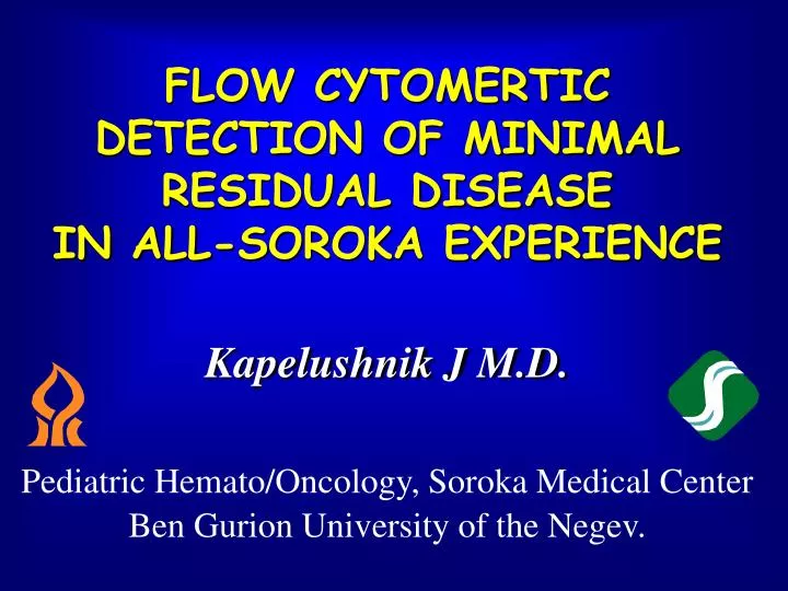
FLOW CYTOMERTIC DETECTION OF MINIMAL RESIDUAL DISEASE IN ALL-SOROKA EXPERIENCE. Kapelushnik J M.D. Pediatric Hemato/Oncology, Soroka Medical Center Ben Gurion University of the Negev. Strategy of Gating. FL2/CD4. FL1/CD8. Lymphocytes. FL2/CD4. FL2/CD4. FL1/CD8. FL4/CD3. FL1/CD8.

E N D
FLOW CYTOMERTIC DETECTION OF MINIMAL RESIDUAL DISEASE IN ALL-SOROKA EXPERIENCE Kapelushnik J M.D. Pediatric Hemato/Oncology, Soroka Medical Center Ben Gurion University of the Negev.
Strategy of Gating FL2/CD4 FL1/CD8 Lymphocytes FL2/CD4 FL2/CD4 FL1/CD8 FL4/CD3 FL1/CD8 SOROKA UNIVERSITY MEDICAL CENTER FLOW CYTOMETRY UNIT
To monitor MRD by flow cytometry, it is essential to characterize antigenic profile of the patient’s leukemic cells at the time of diagnosis and, thereby, to determine the combinations of markers which should be used for detection of residual blasts
To monitor MRD by flow cytometry, it is essential To determine the difference(s) between antigenic properties of leukemic and normal progenitor cells
Flow Cytometric Monitoring of Patients with Acute Leukemia Acute Lymphoblastic Leukemia 2, 12, 20-24, 48-52, 72-75, and 100-104 wk of treatment, after the end of therapy, and 1, 2 and 3 years after the end of chemotherapy
Table. Basic Four-color Staining Protocols for Monitoring of Patients with AL. Staining Protocol (FITC/PE/ECD/PE-Cy5(PerCP) Type of Analyzed Cells Applications* CD45/CD34/CD19/CD33 Lymphoid and myeloid progenitors DIP, BMH, MRD CD20/CD10/CD19/CD45 B cells DIP, BMH, MRD CD45/CD22/CD19/CD24 B cells DIP, BMH, MRD CD31/CD38/CD19/CD45 Activated B cells and plasma cells BMH cyTdT/cyCD79a/CD19/CD45 B cells MRD, BMH CD8/CD4/CD3/CD45 T cells DIP, MRD CD7/CD38/CD45/CD2 T cells, activated T cells DIP, MRD CD8/CD4/cyCD3/CD3 T cells MRD cyTdT/cyCD79a/cyCD3/CD45 T and B cells MRD, BMH HLA-DR/CD11b/CD45/CD33 Myeloid cells, Monocytes DIP, BMH, MRD CD15/CD33/CD45/CD16 Myeloid cells, Monocytes DIP, BMH, MRD CD14/CD13/CD45/CD33 Myeloid cells, Monocytes DIP, MRD CD7/CD117/CD45/CD24 Myeloid cells DIP, MRD CD41/CD64/CD45/CD33 Megakaryoblasts DIP, MRD CD71/GlyA/CD45/CD33 Erythroid cells DIP, MRD cyMPO/cyCD79a/cyCD3/CD45 All types DIP Four Color Staining Protocols for Monitoring of Patients with ALL *Mab used for detection of antigenic markers were correspondingly conjugated with FITC, PE, ECD (PE-TxR), and PE-Cy5 (TC or PerCP. DIP – diagnostic immunophenotyping (i.e. MFC analysis of leukemia cells at the time of diagnosis); BMH – analysis of normal BM hematopoiesis during and after the chemotherapy, MRD – monitoring of MRD. cy – detection of marker in the cytoplasm of cells (intracellular stainig).
HLA-DR/CD11b/CD45/CD33 Myeloid cells, Monocytes DIP, BMH, MRD CD15/CD33/CD45/CD16 Myeloid cells, Monocytes DIP, BMH, MRD CD14/CD13/CD45/CD33 Myeloid cells, Monocytes DIP, MRD CD7/CD117/CD45/CD24 Myeloid cells DIP, MRD CD41/CD64/CD45/CD33 Megakaryoblasts DIP, MRD CD71/GlyA/CD45/CD33 Erythroid cells DIP, MRD cyMPO/cyCD79a/cyCD3/CD45 All types DIP *Mab used for detection of antigenic markers were correspondingly conjugated with FITC, PE, ECD (PE-TxR), and PE-Cy5 (TC or PerCP. DIP – diagnostic immunophenotyping (i.e. MFC analysis of leukemia cells at the time of diagnosis); BMH – analysis of normal BM hematopoiesis during and after the chemotherapy, MRD – monitoring of MRD. cy – detection of marker in the cytoplasm of cells (intracellular stainig).
Study of MRD Detection of Residual Blasts Using Two Staining Protocols (pre-B-ALL; CD10+/CD19+/CD34+ /HLA-DR+ Blasts) I. Flow Cytometry at Diagnosis (35-40% Blasts among WBC) Non-Gated BM Cells Gated (CD45 vs SS) Blast Cells 75% of Blasts 50% of cells in the region of CD45low/SSlow cells (blasts) 75% of Blasts CD10 CD34 SS CD45 CD19 HLA-DR II. Flow Cytometry after Induction Therapy (0.6% Blasts) Non-Gated BM Cells Gated (CD45 vs SS) Blast Cells 2.5% of Blasts 23% of cells in the region 2.5% of Blasts CD10 CD34 SS CD45 CD19 HLA-DR
Residual blasts in bone marrow of patients with ALL 14 cases of ALL Relapse ? CNS Relapse Residual cells (%) Time (wk)
Detection of Blast Cells with Antigenic Properties of Normal Progenitors during Chemotherapy Analysis (CD20 vs. CD10) of Gated CD19+ Cells from Progenitor/Lymphocyte Region (CD45 vs. SS) Patient N.A., B-ALL; 22 wk; Relapse Cluster of CD10+ CD20+ cells; No CD10-CD20+ mature cells Unusually high proportion of cTdT++cCD79a+ Patient A.G., B-ALL; 25 wk; CR Hematologically Normal BM Patient N.A., B-ALL; 49 wk; CR or MRD?
T-ALL (Late Cortical Phenotype) Immunophenotyping at the Time of Diagnosis (~60-70% Blasts) Immunophenotyping at the Time of CNS Relapse (~0.5% Blasts)
AML M7; Diagnosis AML M7; Diagnosis AML M7; CT 8 wk; 1.3% AML M7; CT 6 wk; 0.1% AML M7; CT 13 wk; 1.7% AML M7; CT 17 wk; 13%
Expression of Multidrug Resistance Proteins in Pathologic Cells FL4/ CD33 FL4/ CD33 Mono Myelo Lympho Erythro FL1/ CD45 FL1/ CD45 FL2/ cytoLRP FL2/ cytoMRP2
Conclusions One leukemic cell in 104 normal bone marrow cells can be detected by using flow cytometry. Flow Cytometric detection of residual blasts may be used for the study of early response to chemotherapy. Monitoring of MRD by flow cytometry is useful for early detection of relapse during chemotherapy.
I. Dulman S.Hog Ihia T.Yermiau G. Shubinsky O.Shpilberg A.Moser M. Ben Harush J.Kapelushnik MDR-SOROKA