Download

1 / 72
900 likes | 1.63k Views
Minimal change disease. Introduction. Nephrotic syndrome is kidney disease with proteinuria, hypoalbuminemia, and edema. Nephrotic range proteinuria is 3 grams per day or more Nephrotic syndrome may affect adults and children, of both sexes and of any race. Nephrotic syndrome (NS).

E N D
Introduction Nephrotic syndrome is kidney disease with proteinuria, hypoalbuminemia, and edema. Nephrotic range proteinuria is 3 grams per day or more Nephrotic syndrome may affect adults and children, of both sexes and of any race
Nephrotic syndrome (NS) Classification • Nephrotic syndrome can be primary, being a disease specific to the kidneys, or it can be secondary, being a renal manifestation of a systemic general illness • In all cases, injury to glomeruli is an essential feature
Primary causes of nephrotic syndrome (NS) Include, in approximate order of frequency : • Minimal-change nephropathy • Focal glomerulosclerosis • Membranous nephropathy • Hereditary nephropathies
Secondary causes of NS Include, again in order of approximate frequency : • Diabetes mellitus • Lupus erythematosus • Amyloidosis and paraproteinemias • Viral infections (eg, hepatitis B, hepatitis C, human immunodeficiency virus [HIV] ) • Preeclampsia
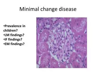
Minimal change disease • Most common cause of the nephrotic syndrome (NS) in children • ~10-15% of NS in adults, third most common after MN and FSGS • More common in Hispanics, Asians, Arabs and Caucasians • Clinical and pathologicalentity defined by selective proteinuria and hypoalbuminemiathat occurs in the absence of • cellular glomerular infiltratesor • immunoglobulin deposits
NS in infancy and childhood is an important entity • A study from New Zealand found the incidence of nephrotic syndrome to be almost 20 cases per million children under age 15 years 1 • In specific populations, such as those of Finnish or Mennonite origin, congenital nephrotic syndrome may occur in 1 in 10,000 or 1 in 500 births, respectively 2 • 1. J Paediatr Child Health. May 2007;43(5):337-41 • 2. Pediatr Nephrol. Dec 2004;19(12):1313-8
According to the International Study of Kidney Diseases in Childhood (ISKDC) • 84.5% of all children with primary nephrotic syndrome have minimal-change nephrotic syndrome (MCNS) • 9.5% have focal segmental glomerulosclerosis (FSGS) • 2.5% have mesangial proliferation, and • 3.5% have membranous nephropathy or another cause of the disease 1,2 • MCNS remains the most important cause of chronic renal disease in children • 1. Kidney Int. Dec 1981;20(6):765-71 • 2. J Pediatr. Apr 1981;98(4):561-4
Pathophysiology • Primary urine is formed through the filtration of plasma fluid across the glomerular barrier; the glomerular filtration rate (GFR) is 125 mL/min • The plasma flow rate (Qp) is close to 700 mL/min, with the filtration fraction being 20% • The concentration of albumin in serum is 40 g/L, while the estimated concentration of albumin in primary urine is 4 mg/L, or 0.1% of its concentration in plasma GBM = glomerular basement membrane; Endo = fenestrated endothelial cells; ESL = endothelial cell surface layer (often referred to as the glycocalyx).
The barriers that keep protein and blood cells out of the urine. These are the endothelial cell, basement membrane and epithelial cell (podocyte). The epithelial cell (podocyte) seems to be most important. Injury to these barriers causes proteinuria and hematuria
Pathophysiology (contd.) • The glomerular structural changes that may cause proteinuria are - (1) damage to the endothelial surface, - (2) damage to the glomerular basement membrane, - and/or (3) damage of the podocytes • In congenital nephrotic syndrome, the gene for nephrin, a protein of the filtration slit, is mutated, leading to nephrotic syndrome in infancy
Pathophysiology (contd.) • Albuminuria alone may occur, or, with greater injury, leakage of all plasma proteins, (ie, proteinuria) may take place • Proteinuria that is more than 85% albumin is selective proteinuria • In minimal-change nephropathy, proteinuria is selective
Pathogenesis of edema • An increase in glomerular permeability leads to albuminuria and eventually to hypoalbuminemia • In turn, hypoalbuminemia lowers the plasma colloid osmotic pressure, causing greater transcapillary filtration of water throughout the body and thus the development of edema • A reduction in plasma volume, with a secondary increase of sodium and water retention by the kidneys
Metabolic consequences of proteinuria • levels of serum lipids are usually elevated • The loss of antithrombin III and plasminogen and increase in clotting factors, especially factors I, VII, VIII, and X, increases the risk for venous thrombosis and pulmonary embolism • Hypovitaminosis D - malabsorption of Ca++ • Lower the patient's resistance to infections and increase the risk of sepsis and peritonitis
Immunofluorescence Microscopy www.gamewood.net/rnet/renalpath/noimcx.jpg
The glomerular capillary wall Normal MCD Van den Berg, Weening, Clinical Science (2004) 107, 125–136
Pathogenesis - “Intrinsic factor” • Genetic basis for hereditary NS • NS of the Finnish type • Autosomal-recessive steroid-resistant NS • Familial forms of FSGS • Diffuse mesangila sclerosis associated with Denys-Drash syndrome and with Frasier syndrome • NS associated with nail-patella syndrome • Help elucidate molecular aspect of FSGS • Not clear for MCD
Molecular anatomy of the podocyte foot process cytoskeleton Nature Genetics24, 333 - 335 (2000)
Pathogenesis – extrinsic factor, better explanation for MCD • Clinical Observations - Shalhoub’s hypothesis • MCD frequently remits with measles infection • Corticosteroids and alkylating drugs cause a remission • Association of MCD with Hodgkin disease • Experimental Observations • T cell hybridoma (Koyama KI 1991 (40): 453-460) • Removal of glomerular permeability factor leads to normal kidney (Ali Transplantation 1994 Oct 15;58(7):849-52) • “circulating factor” • possible link between T-cell response and glomerular disease
MCD is a disorder of T cells • T-cells release a cytokine that injures the glomerular epithelial foot processes • This leads to a decreased synthesis of polyanions • The polyanions constitute the normal charge barrier to the filtration of macromolecules, such as albumin • When the polyanions are damaged, leakage of albumin follows • The identity of this circulating permeability factor is uncertain, although it is postulated that it may be hemopexin
Some of the cytokines that have been studied in MCD are interleukin-12 (IL-12) and interleukin-4 (IL-4) • IL-12 levels have been found to be elevated in peripheral blood monocytes during the active phase and normalized during remission • Interleukin-18 (IL-18) can synergize with IL-12 to selectively increase the production of vascular permeability factor from T cells • In addition, levels of IL-4 and CD23 (a receptor for immunoglobulin E [IgE] 1 have been found to be elevated in peripheral blood lymphocytes • 1. Am J Med Sci. Oct 2009;338(4):264-7
Synaptopodin is a proline-rich protein intimately associated with actin microfilaments present in the foot processes of podocytes • Greater synaptopodin expression in podocytes is associated with a significantly better response to steroid therapy • Interleukin-13 (IL-13) has been implicated in the pathogenesis of MCD. • IL-13 genetic polymorphisms correlate with the long-term outcome of MCD. • IL-13 overexpression can cause podocyte foot process fusion and proteinuria 1 • 1. May 2007;18(5):1476-85
Overexpression of Interleukin-13 Induces Minimal-Change–Like Nephropathy in Rats • Background • MCD may be a T cell dependent disorder that results in glomerular podocyte dysfunction • Th2 cytokine bias in patients with MCD • MCD associated with atopy and allergy • Relapse MCD with elevated IL-4 and IL-13 • Association between MCD and Hodgkins’s disease • IL-13 known to be an autocrine growth factorfor the Reed-Sternberg
Hypothesis IL-13 may play an important role in the development ofproteinuria in MCNS by exerting a direct effect on podocytes,acting through the IL-13 receptors on the podocyte cell surface,initiating certain signaling pathways that eventually lead tochanges in the expression of podocyte-related proteins (nephrin, podocin, and dystroglycan) IL-13 transfected mouse was used as a model
Comparison of control, IL-13-transfected mouse at experiment end (day 70) Yellow = p <0.001 vs control Red = p<0.001 vs control and Grp 1
Histopathologic features on day 70 at killing(A) Glomerulus of IL-13–transfected rat showing no significant histologic changes (periodic acid-Schiff stain). (B) Glomerulus of IL-13–transfected rat showing fusion of podocyte foot processes (arrows). (C) Glomerulus of control rat showing normal individual podocyte foot processes along the glomerular basement membrane (GBM; arrows).
Immunofluorescence staining of glomeruli for protein expression of nephrin, podocin, dystroglycan, and synaptopodin Control IL-13 infected nephrin podocin dystroglycan synaptopodin
Summary • IL-13-transfected rats • Developed minimal change like GN, as evidence by LM and EM changes • decrease in the expression of nephrin, podocin,and dystroglycan associated with increased urinary albumin excretion and podocytefoot process effacement • suggesting that these proteins areessential in maintaining the filtration barrier, thus controllingglomerular permeability • decrease was not due to loss ofpodocytes -
In patients who develop acute renal failure, endothelin 1 expression is greater in the glomeruli, vessels, and tubules than in the nonacute renal failure group • The glomerular epithelial cells (podocytes) and the slit diaphragm connecting the podocyte foot processes play a primary role in the development of proteinuria • Nephrin is a major component of the slit diaphragm. The slit diaphragm is often missing in MC nephrotic syndrome (MCD) kidneys • The role of nephrin and the slit diaphragm in MCD is not known. However, genetic variants of a glomerular filter protein may play a role in some patients with MCD
Izzedine et al found a lack of glomerular dysferlin expression associated with minimal-change nephropathy in a patient with limb-girdle muscular dystrophy type 2B. 1 • In the same study, 2 of 3 other patients with dysferlinopathy had microalbuminuria • Although a multitude of studies have been published, the mechanism by which T cells increase glomerular permeability has remained unproven • 1. Am J Kidney Dis. Jul 2006;48(1):143-50
Frequency • United States - In preadolescents, minimal-change nephrotic syndrome (MCNS) makes up 85-95% of all cases of nephrotic syndrome • In adolescents and young adults, the prevalence is 50%, while in adults, MCNS accounts for 10-15% of primary nephrotic syndrome cases. • The incidence of nephrotic syndrome is 2-7 new cases annually per 100,000 children, and the prevalence is 15 cases per 100,000 children • Asians may be at increased risk.
Incidence of important causes of nephrotic syndrome, in number per million population • The left panel shows systemic causes, and the right panel lists primary renal diseases that can cause nephrotic syndrome. • fgs = focal glomerulosclerosis, • MN = membranous nephropathy, • min change = minimal-change nephropathy Clin J Am Soc Nephrol. May 2006;1(3):483-7 Nephrol Dial Transplant. 2007;22:1608-1618
Sex / Age • It is found twice as frequently in boys than in girls • The frequency is the same between the sexes in adults • The incidence peaks in children aged 2 years, with approximately 80% being younger than 6 years at the time of diagnosis • In adults, the mean age of onset is 40 years.
A schema of the average patient ages associated with various common forms of nephrotic syndrome
History • Edema may be preceded by an upper respiratory tract infection, an allergic reaction to a bee sting, or the use of certain drugs or malignancies. • Facial edema is noted first. • Malaise and easy fatigability can occur. • Weight gain often is an additional feature. • The patient also may present with the following: • Hypovolemia • Hypertension • Thromboembolism • Infection
Physical • BP - usually is normal in childrenbut may be elevated in adults • Dependent edema is the most prominent sign. The retina has a wet appearance. Subungual edema with horizontal lines (called Muehrcke lines) also may occur. • Hernias may be found, and the elasticity of the ears may be decreased. • Heavy proteinuria - leads to a state of protein depletion with muscle wasting, thinning of the skin, and growth failure • Pleural and ascitic fluid can accumulate. Rarely, cellulitis, peritonitis, or pneumonia • Children may have growth failure.
Causes • Almost all cases are idiopathic, but a small percentage of cases (approximately 10-20%) may have an identifiable cause • Causes may include the following: Secondary • Drugs - Nonsteroidal anti-inflammatory drugs (NSAIDs), rifampin, interferon, ampicillin/penicillin, trimethadione, mercury-containing cosmetic skin cream • Toxins - Mercury, lithium, bee stings, fire coral exposure • Infection - Infectious mononucleosis, HIV, immunization • Tumor - Hodgkin lymphoma (most commonly), carcinoma, other lymphoproliferative diseases • Posthematopoietic stem cell transplant
Laboratory Studies • Urine analysis - profound proteinuria and oval fat bodies observed. • In children, the critical level for diagnosis is more than 40 mg/h/m2. • In adults, the threshold is more than 3.5 g/d/1.73 m2. • Albumin-to-creatinine concentration ratio is in excess of 5. • Urine specific gravity is high because of proteinuria. • A 24-hour urine is obtained for protein and creatinine clearance. • Hypoalbuminemia - Nephrotic syndrome in children is defined by a serum albumin of less than 2.5 g/dL. • Hyperlipidemia also is a feature of a nephrotic state. • Renal function usually is normal except in cases of ARF • Hyponatremia often is observed, • Elevated hemoglobin and hematocrit
Imaging Studies - Renal sonogram is normal. • Procedures • Because of the high prevalence of MCD in children with nephrotic syndrome, an empiric trial of corticosteroids commonly is the first step in therapy • Renal biopsy typically is performed only in resistant cases • Generally, if proteinuria remains after 2 relapses or courses of steroids, a tissue diagnosis should be made before starting cytotoxic or immunosuppressive therapy
Medical Care • Corticosteroids are the treatment of choice, leading to complete remission of proteinuria in most cases • Approximately 90% of children respond within 2 weeks to prednisone at a dose of 60 mg/msq/d. • The treatment is continued for another 6 weeks, at lower doses of prednisone, after the remission of proteinuria. • In some children, proteinuria fails to clear by 6-8 weeks, and performing a renal biopsy may be useful to determine if another process may be present
Adults respond more slowly than children • A response in up to 80-90% in adolescents and adults • The time to remission is up to 16 weeks. If patients are steroid-resistant or they relapse frequently, a trial of immunosuppressants is given • Immunosuppressants - cyclophosphamide and chlorambucil • Cyclosporine is considered to be an acceptable drug for maintenance therapy in patients with frequent relapses and steroid dependency. However, it is less efficacious than cyclophosphamide at maintaining sustained remission
Response of patients to steroids is used to divide patients into various groups. • Complete remission: This is defined as complete resolution of proteinuria for at least 3-5 consecutive days. • Partial remission: This is defined as a reduction in the degree of proteinuria without complete clearing. • Relapse: This is defined as a reoccurrence of proteinuria for at least 3-5 consecutive days.
Because MCNS accounts for 90% of all cases of idiopathic nephrotic syndrome in children, steroids are started empirically. A biopsy is performed only in those cases where no remission occurs • In comparison, a biopsy is performed in all adults before the initiation of treatment. Adults tend to respond more slowly, with more than 25% taking as long as 12-16 weeks to undergo complete remission • Initial regimen in adults consists of oral prednisone in a daily dosage of 1 mg/kg of body weight for 8-16 weeks (or for 1 wk after remission has been induced). The patient is then placed on an alternate-day single-dose (1 mg/kg) regimen to minimize the incidence of adverse effects. • If proteinuria disappears or is reduced to a very low level, high-dose alternate-day therapy is continued for several weeks to 1 month and then slowly tapered over several months in an attempt to reduce the likelihood of relapse