Download
1 / 1
10 likes | 135 Views
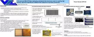
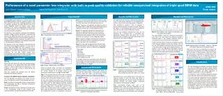
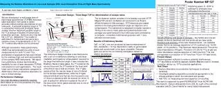
Novel 3-D Sample Plate using Monolithic Capture Media in Collimated-Hole Structures for Interfacing High Capacity Separations with MALDI-TOF. 6. 1479 select. 25000. 20000. 15000. Area 1479. C. B. A. 1479. 927. 10000. 5000. 1725. 800. 0. 0. 2000. 4000. 6000. 8000. 10000. fmol.

E N D
Novel 3-D Sample Plate using Monolithic Capture Media in Collimated-Hole Structures for Interfacing High Capacity Separations with MALDI-TOF 6 1479 select 25000 20000 15000 Area 1479 C B A 1479 927 10000 5000 1725 800 0 0 2000 4000 6000 8000 10000 fmol 600 TIC 1300-2400 (x1000) 400 EIC 900-2400Da EIC 3 individual peptides EIC matrix 200 0 0 1000 2000 3000 4000 5000 6000 7000 8000 fmol Poster Number TP 060 Stephen Hattan, Marvin Vestal Virgin Instruments Corporation Sudbury, Massachusetts USA 13x increase 3 Overview/Introduction: -Novel MALDI targets are being developed that use collimated hole structures (CHS) combined with monolithic chromatography media to enable capture & concentration of sample and serve as a direct interface between the mass spectrometer and different separation schemes -HPLC, electrophoresis and tissue imaging- -Plate construction, sample deposition (LC) and sample elution are presented -Results of capacity and LC interface at 15 and 50 μL/min separation speeds are presented 8 1 • Figure 3: Prototype LC deposition System • X Y control of solvent capillary • Tip seals with plate surface • column pressure forces column effluent through discrete locations on CHS plates. 32x increase Figure 8: Four Identical, 25 min, 50μL/min LC separation on single CHS plateConditions: 50x1mm, 200Å, C-18 5μm, Higgins Anal., 0-45% ACN (25 min) 50μL/min flow, dwell time/spot 10s -overlay of EIC from 12 mid-intensity range peptides plotted as ratio against1042internalstandardand show and average relative standard deviation of 22%. -5pmol of BSA digest run in series and spotted on a single 675 well CHS plate (Fig. 1 top middle) -separations conditions represent a 50-100X increase in flow rate/sample load and capture over conventional LC-MALDI schemes performed on 2D plates. -relative large sample load, high speed, reproducible separation afforded by CHS plates. Figure 6: Comparison with conventional 2D plate: -Identical 1hr separation of 1pmol BSA at 15uL/min flow spotted at 5s intervals on a 2D surface and 20s intervals on a CHS plate. -Left frames shows the EIC and max-intensity spectrum of 1479Da peptide from each run (CHS top:2D bottom). -Right frames are blow-ups of the EIC region of interest. -Right top: raw signal intensity 13x increase in intensity -Right bottom: signal intensity normalized against a 1042Da internal standard (matrix addition) demonstrates 32x increase in signal intensity Based dwell time and spot dimensions a 36x increase in signal was calculated therefore the above results indicate near 100% sample recovery. 4 ~2x2” 100 well 5x5” 675 well Std. 384 well Figure 4: Prototype Elution System -uses pressure on reservoir of eluent residing on one side of the plate to force the liquid through all holes simultaneously. -enclosed chamber with exhaust fan on opposite side of plate enhances solvent evaporation and matrix crystallization. μ- Channel 2x2” Figure 1: CHS plates: Row 1: 100, 675 and 384 wells plate of 1.5, 3 and 10 mm thickness respectively constructed in metal and plastic designed for modes of discrete sample deposition such as LC or robotic and hand spotting. Row two shows glass μ-channel CHS plate (25μm holes) designed for interface with gel and tissue applications. Conclusions: -Novel 3-dimensional MALDI target that captures and concentrates peptide/protein sample and serves as a direct interface between separation and mass spectrometer -Plates allow for higher load capacity (potentially 100s μg to mg quantity) and higher flow (100s μL- mL/min) separations that should enable dramatic increase in the detection levels of lower concentration proteins/peptides in complex proteomic mixtures. -Plates are designed to be reusable with current prototypes easily withstanding 25-50 analyses (like typical LC columns). -Presentation focuses on the reversed phase LC-MALDI interface because of its near ubiquitous capture of peptide material; however, plates focused on affinity capture (ie. glyco-capture) and studies focused on direct interface of Gel and tissue sample preparation schemes with MALDI are underway. Acknowledgement: Recognition and thanks is deserved for the entire Virgin Instruments staff for significant contributions to this project; Christina Vestal, Kevin Hayden, Roger Voyer, Joseph Fitzpatrick, Joseph Valentine “as in Valentine’s Day”, George Mills, Steve Gabeler, Matthew Gabeler and Mark Dahl. Funding: This work was supported by SBIR Grant GM079833 7 10fmol-10pmol dilution series different sample Conc. loaded in identical volumes 2 5 10fmol-7.5pmol conc. series identical sample loaded in different volumes • Figure 5 LC Results: • 3D plots of signal intensity as function of surface location from 1pmol BSA digest LC separation Conditions: 0.5 mm column ID C-18 (eksigent), 15uL/min, 5-45%ACN gradient (50min), dwell time 30s/spot 10x10 CHS plate (Fig. 1 top left) • -Frame A: extracted ion chromatogram (EIC) all peptides from 900-2400 Da. • -Frame B: EIC of three individual peptides (927,1479,1725 Da) • -Frame C: EIC of 379Da ACCH matrix dimer. • Figure demonstrates the ability of the CHS plates to capture/concentrate sample without loss of chromatographic resolution and uniform sample elution. Figure 2: Plate Construction: -styrene/divinylbenzene based polymers for reversed phase capture media of protein/peptides -monomer solutions are injected into tetrafluoroethylene molds -polymerization is thermally initiated -Excess polymer is shaved to a level coincident with the plate substrate (metal, plastic, glass). -ratio and constitution of dilution solvent(porogens) are used to tailor pore-size/flow properties -Sample application/analysis occur on the polymer surface. • Figure 7: Capacity study: • plots from different loading schemes aimed at determining capacity of CHS plate with ~5μL void volume. • Plot A: dilution series of different conc. loaded in some volume as not overwhelm the void volume of well shows ~ 8pmol load capacity limited primarily by analyte/matrix ratio/ ionization efficiency • Plot B Same sample different load volume --void volume exceeded-- Limited by binding capacity of CHS wells ~2.5 pmol of digested protein. • Binding capacity will vary with well dimensions


![[INSERT] Poster number (eg P324, or OP212). Title of your poster.](https://cdn3.slideserve.com/6811451/slide1-dt.jpg)


![Prepare 210-060 Exam with Actual 210-060 Dumps [PDF]](https://cdn4.slideserve.com/7889925/cisco-dt.jpg)