Download
1 / 50
520 likes | 562 Views
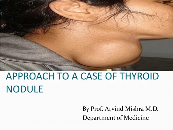
Approach to a thyroid nodule. Anatomy of the Thyroid Gland. Follicles: the Functional Units of the Thyroid Gland. Follicles Are the Sites Where Key Thyroid Elements Function: Thyroglobulin (Tg) Tyrosine Iodine Thyroxine (T 4 ) Triiodotyrosine (T 3 ). Thyroid Hormones.

E N D
Follicles: the Functional Units of the Thyroid Gland Follicles Are the Sites Where Key Thyroid Elements Function: • Thyroglobulin (Tg) • Tyrosine • Iodine • Thyroxine (T4) • Triiodotyrosine (T3)
Thyroid Hormones Thyroid pro-hormone is stored as thryoglobulin as an extracellular colloid T3 and T4 can cross lipid membranes readily (secretion and uptake) T3 and T4 are small, hydrophobic and circulate bound to Thyroxine-binding globulin (TBG)
Approach • Clinical. • Biochemical. • Radiological. • Histopathological.
Clincal • History taking. • Physical examination.
History • Profile. • Mass in ant. neck (onset, duration,pain, course, trauma….) • Assessment of function( Symptoms of thyrotxicosis or hypothyroidism) • Risk factors for malignancy. • Review of Systems, medical hx., past hx, drug hx, social hx.
Symptoms of thyrotoxicosis • nervousness, tremors, sweating, heat intolerance, palpitations, wt loss despite normal or increased appetite, amenorrhea, weakness.
Hypothyroidism • Lethargy, hoarseness, hearing loss, thick and dry skin, constipation, cold intolerance, stiff gate, weight gain.
Risk factors for malignancy Age sex occupation family hx Painless Hoarseness Hx of irradiation hard, LN enlargement residency,...............etc.
Physical Exam • Swelling in the anatomical site of thyroid. • Moves with swallowing
Goitre • Diffuse • Nodular -Solitary nodule -Multinodular goitre
Anatomical dx includes retrosternal extension extension below sternocledomastoid
Solitary Noudule • Neoplastic • Non neoplastic
Non Neoplastic • Cyst: degenerative, Hemorrhagic, Hydatid… Surgery is indicated after second recurrence. • Solid: Part of Multinodular Goiter.
Neoplastic • Benign: Follicular adenoma • Malignant: Wide spectrum of behaviour
Papillary Ca • Most common, Best prognosis • 10 year survival around 85 % • At younger age group. • Spreads by lymphatics. • Can be multifocal. • Can be familial. • Usually sensitive to RAI
Follicular Ca • 10 y survival around 60 %. • Associated with iodine deficiency. • Usually monofocal. • Haematogenous spread. • Diagnosed by capsular and vascular infiltration. • Sensitive to RAI.
Medullary Ca • From Parafollicular cells. • 10 year survival 25-30% • Can be Familial or Sporadic. • Can be part of MEN 2. • Does not uptake RAI.
Anaplastic • Around 1 % • Very aggressive tumor. • The worst prognosis • Survival is usually less than 6 months
Fibrolymphovasclar tumors • Haemangioma, Lymphoma, Fibroma,….. • Secondary Metastases.
Biochemical • Thyroid function tests: T3, T4, TSH. • Antithyroid Antibodies: antithyroglobulin, antimicrosomal antibodies.
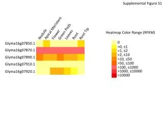
Imaging Assessment • Ultrasound. • Computerized tomographic scan. • Magnetic resonance scan. • Radioactive Iodine scan.
Pathological Dx • Fine Needle Aspiration. • Surgery for definitive biopsy.
Ultrasound • One nodule or more • Cystic or solid • Presence or abecence of features of malignancy • Cervical LN enlargement
Features of malignancy in U/S • Microcalcification. • Hypoechoeic nodules. • Increased vascularity. • Interupted hallo sign
U/S guided FNA • Prefered if - > 50 % cystic leision. - located posteriorly.
Serum Thyroglobulin • Increases in most thyroid pathologies. • Not specific as a diagnostic tool. • For follow up only.
Serum Calcitonin • Contraversy about its importance as a diagnostic tool. • if >100 pg/ml can suggest medullary Ca.
Benign FNA • Risk of false neg. Up to 5%. papable >U/S guided(0.6%). • Repeat examination or U/S 6-18 m interval • Growth>20%,or more than 2mm in two dimensionsrepeat FNA preferably U/S guided.
Medical Treatment • No data to suggest that TSH suppression will cause a change in thyroid nodule size in Iodine sufficient area. • Not recomended.
Children • Should be evaluated as adults.
Pregnancy • Thyroid scan should be delayed till delivery. • If operation is to be done 12-24wks GA. • After that should be postponed till delivery. • (studies:delay less than one year will not affect the eventual prognosis)
Treatment • Goals: 1-to remove the primary tumour and its local extension. 2-to minimize treatment related morbidity. 3-to permit accurate staging. 4-fascilitate postop. Radioactive Iodine ttt. 5-fascilitate long term postop. Surveilance 6-minimize disease reccurence and mets.
Total Thyroidectomy • 1- FNA papillary,medullary. 2- nodule > 4cm and atypia. 3- hx. Of irradiation or positive family hx. 4- bilateral nodules. 5- regional LN or distant metastases. 6- patient preferance for one stage. 7- relative indicationage >45
Lobectomy • Soitary nodule+indetermined pathology FNA+ patient preferance.
Central LN Dissection • CLN are most common site of recurrence. • Routine CLN dissection is indicated in medullary Ca., no consensus in papillary Ca.
Lateral Neck Dissection Levels II,III,IV and V Done only with biopsy proven metastases after clinical or sonographic suspicion
Completion Thyroidectomy • To allow resection of multicentric disease. • Allow radioactive Iodine diagnostic scan and treatment. • Studies:same surgical risk as one stage surgery. • (small tumours<1cm,intrathyroid,node neg.,low risk group) can be managed without completion.
Postoperative Radioactive Iodine Ablation • Prepared with L-thyroxin withdrawal for 4 wks,or replace it with T3 for 2-4 wks then withdraw it for 2 wks. • TSH > 30, to increase avidity. • The minimal activity should be used 30-100 mci. • Higher dose 100-200, in residual disease or aggressive pathology(tall cell,columnar,insular)
Recombinant human thyrotropin(rhTSH) can be used in patients who cannot tolerate stopping thyroxin. • Needs stopping thyroxin for one day only. • Approved in Europe but still not in USA.
Whole body scan • Usually done one week after ablation therapy. • 10-26% metastatic foci.
External Beam Radiotherapy • Indications - age > 45 and extrathyroid extension and high likelyhood of microscopic residual tumour. -gross residual and further surgery or radioactive iodine treatment is ineffective.
Chemotherapy • NO role for chemotherapy in differentiated thyroid Ca. • Some studies:Adriamycin can act as a radiation sensitizer for external beam radiotherapy.
TSH Suppression Therapy • Differntiated thyroid Ca have TSH receptors on cellular membrane. • High risk patients < 0.1 mu/l • Low risk patients 0.1 - 0.5 mu/l
Follow Up • Every 6-12 months. • Physical examination and cervical U/S • Thyroglobulin and calcitonin. • In borderline Tgn stimulation by withdrawing thyroxin or rhTSH. • If positive whole body scan