Download
1 / 46
600 likes | 1.02k Views
Macromolecules in the cell. 513341 BIOCHEMISTRY I Chapter 2 Amino acids and Primary structure. Dr. PORNTIP CHAIMANEE Chemistry Department Faculty of Science Silpakorn University. Amino acids. 1. Amino acids structure 2. Asymmetry of amino acids

E N D
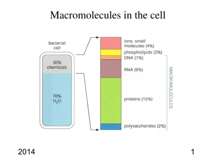
513341 BIOCHEMISTRY I Chapter 2 Amino acids and Primary structure Dr. PORNTIP CHAIMANEE Chemistry Department Faculty of Science Silpakorn University
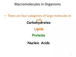
Amino acids 1. Amino acids structure 2. Asymmetry of amino acids 3. Acid and Base properties of amino acids
Three classes of amino acids 1. Non polar R groups 2. Uncharged polar R groups 3. Charges polar R group
Stereochemistry of Amino Acids D,L (Fischer)Nomenclature • Horizontal substituents are pointing towards you • Vertical substituents are pointing away • Carbon in highest oxidation state is at the top • D if the non-hydrogen group is to the right • For glycine both horizontal groups are H - achiral L-Alanine D-Alanine
Stereochemistry of Amino Acids D,L (Fischer) Nomenclature • D,L-nomenclature is based on D- and L-glyceraldehyde • L-amino acids predominate in nature
Acid and base properties of amino acids Amphoteric @ All amino acids have three ionizable groups except glycine @ carboxyl at Ca with pKa between 1.8-2.8 @ amino group with pKa between 8.8-10.6 @ side chain R- groups
Acid-Base ChemistryAmino acids are weak polyprotic acids It has both anionic and cationic properties
The pH at which amino acid has no net charge is called the isoelectric point (pI) Titration curve of glycine Sodium hydroxide glycine
Monopositive Isoelectric Mononegative Dinegative (Cation) (Zwitterion) (Zwitterion) (Anion) Electrical properties and titration curve of Glutamic acid Isoelectric point (pI) for amino acids with ionizable side chains: Take average pKa for the two ionizations involving the neutral (net charge of zero) species. pI of Glu = (2.2 + 4.3)/2 = 3.3
Sample Calculation What is the pH of a glutamic acid solution if the alpha carboxyl is 1/4 dissociated? • Glu+ is dissociated 1/4 so Glu0is 1/4 and Glu+(what’s left) is 3/4. • Use the Henderson-Hassebalch Equation : pH = pKa of COOH+ log [A-]/[HA] pH = pKa of COOH + log [Glu+]/[Glu+] pH = 2 + log (0.25/0.75) pH = 2 + (-0.477) pH = 1.523
Dipositive Monopositive Isoelectric Mononegative (Cation) (Zwitterion) (Zwitterion) (Anion) Electrical properties and titration curve of Lysine a b g pKe-NH2 pK a-NH2 pK a-COOH d e pI of Lys = (9.2 + 10.8)/2 = 10
Another Sample Calculation At pH 9.5, what percentage of the alpha- and epsilon- amino groups of a 1 mM lysine solution are not protonated? For each amino group: [NH2] + [NH3+] = 1 x 10⁻³ For the a-amino group 9.5 = 9.0 + log[NH2]/[NH3+] 3.16 = [NH2]/[NH3+] So [NH3+] = 0.24mM and [NH2] = 0.76mM 76% is present as NH2 For the e-amino group 9.5 = 10.5 + log10[NH2]/[NH3+] 0.1 = [NH2]/[NH3+] So [NH3+] = 0.91mM and [NH2] = 0.09mM 9% is present as NH2 At pH 9.5 the ratio of a-NH2 to the e-NH2 is 8.4:1 This is the kind of thing that’s useful to know if you want to do chemistry specifically at the a-amino group of a peptide The Henderson-Hasselbalch equation allows calculation of the ratio of a weak acid and its conjugate base at any pH
His0 His- His2+ His+ Electrical properties and titration curve of histidine pI of His = (6.0 + 9.17)/2
The pK values of the a-carboxyl, a-amino groups and side chains found in the individual amino acids pI (isoelectric point) 6.2 5.9 5.9 5.7 5.9 2.8 3.2 7.6 5.0 5.6 9.7 10.7
Reactions of Amino Acids • Amino groups form Schiff ‘s bases and amides • Carboxyl groups form amides & esters • Some side chains show unique reactivities – Cys residues can form disulfides and can be easily alkylated – Cys His Asp Glu common metal-binding ligands
Chemical properties of amino acids NINHYDRIN REACTION a - amino acid + Ninhydrin Violet solution + 3
Spectroscopic properties UV Absorbance Spectra of Aromatic Amino Acids • All amino acids absorb in infrared region • Only Phe, Tyr, and Trp absorb UV • Absorbance at 280 nm is a good diagnostic device for amino acids
Biological active amino acid derivatives If the acid is removed , it is converted to Dopamine Glutamic acid COOH Dihydroxyphenylalanine GABA COOH Histidine Thyroxine Histamine
Formation of a Peptide Peptides • Short polymers of amino acids • Each unit is called a residue • 2 residues - dipeptide • 3 residues - tripeptide • 12-20 residues - oligopeptide • many - polypeptide Glycylalanine
Propertiesofpeptide Direction of peptide chain amino or N-terminus carboxyl or C-terminus Peptides • Short polymers of amino acids • Each unit is called a residue • 2 residues - dipeptide • 3 residues - tripeptide • 12-20 residues - oligopeptide • many - polypeptide
Primary structure or sequence SEQUENCE The single- and three- letters codes for amino terminal of a primary sequence
Many peptides are biologically active Two amino acid difference at position 3 and 8 !!!!! Oxytocin -stimulates uterine contractions during labor Vasopressin -Antidiuretic *adjust water reabsorbed by the kidney
Primary structure or sequence The single- and three- letters codes for amino terminal of a primary sequence
Primary Structure of Bovine Insulin Hormone insulin that regulates sugar levels in blood. First protein to be fully sequenced (by Fred Sanger in 1953). For this, he won his first Nobel Prize. Insulin compose with two chains (A and B) Amino acid sequence of human insulin is very similar to that from cows and pigs (only amino acids at 8,9,10 (A) and 30 (B) differ )
Ion-Exchange Chromatography Separation of amino acid mixtures by chromatography(Ion exchange or High-performance liquid chromatography) Amino acids Amino acids
Identification of N and C-Terminal Residue • N-terminal analysis: • FDNB • Edman degradation using phenylisothiocyanate and derivatives are phenylthiohydantions (PTH) • C-terminal analysis • (1)Enzymatic analysis (C-terminus-specific exopeptidase ,carboxypeptidase). • – Carboxypeptidase A cleaves any residue except Pro, Arg, and Lys • – Carboxypeptidase B (hog pancreas) only works on Arg and Lys • (2) done by hydrazinolysis (reaction with anhydrous hydrazine in presence of mildly acidic ion exchange resin) NO2 (trifluoroacetic acid) (PTC) (PTH) and identified by HPLC
Fragmentation of the chains Enzymatic fragmentation– trypsin, chymotrypsin, clostripain, staphylococcal protease
Fragmentation of the chains Chemical Fragmentation :Cyanogen bromide CNBr acts only on methionine residues CNBr is useful because proteins usually have only a few Met residues
Reconstructing the Sequence • Use two or more fragmentation agents in separate fragmentation experiments • Sequence all the peptides produced (usually by Edman degradation) • Compare and align overlapping peptide sequences to learn the sequence of the original polypeptide chain Compare cleavage by trypsin and staphylococcal protease on a typical peptide: • Trypsin cleavage: A-E-F-S-G-I-T-P-K L-V-G-K • Staphylococcal protease: F-S-G-I-T-P-K L-V-G-K-A-E • The correct overlap of fragments: L-V-G-K A-E-F-S-G-I-T-P-K L-V-G-K-A-E F-S-G-I-T-P-K • Correct sequence: L-V-G-K-A-E-F-S-G-I-T-P-K
A molecular clock • Plot the number of changes in amino-acids between the same protein in different species (such as cytochrome C) against the time since the species diverged • Gives a straight line - so evolution of a protein sequence proceeds at a constant rate and therefore can be used as a clock • Calibration of the clock for specific protein families would ensure the dating of biological events not present in the fossil record and would imply that changes are non-adaptive due to their independence of the selective constraints