Download
1 / 24
240 likes | 246 Views
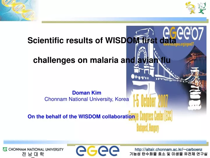
Scientific results of WISDOM first data challenges on malaria and avian flu. Doman Kim Chonnam National University, Korea. On the behalf of the WISDOM collaboration. Searching for new drugs. Drug development is a long (10-12 years) and expensive (~800 MDollars) process

E N D
Scientific results of WISDOM first data challenges on malaria and avian flu Doman Kim Chonnam National University, Korea On the behalf of the WISDOM collaboration
Searching for new drugs Drug development is a long (10-12 years) and expensive (~800 MDollars) process In silico drug discovery opens new perspectives to speed it up and reduce its cost From Dr. Vincent Breton
A first step towards in silico drug discovery: virtual screening In silico virtual screening Starting from millions of compounds, select a handful of compounds for in vitro testing Very computationally intensive but potentially much cheaper and time effective than typical in vitro testing From Dr. Vincent Breton
Molecular docking Millions Molecular dynamics 5000 AMBER Re-ranking MMPBSA-GBSA Ligand CHIMERA Amber 4 H bonds Complex visualization 180 Ligand Catalytic aspartic residues Catalytic aspartic residues Ligand 2 Hydrogen Bonds Catalytic aspartic residues In vitro tests 30 WET LABORATORY Wisdom I workflowfor malaria inhibitor development FLEXX AUTODOCK From Ana & Vinod
Areas where malaria transmission occurs Areas with limited risk No malaria Inhibition of Plasmepsin II
Plasmepsin II Hemoglobin (Hb) Plasmepsins I, II, IV and HAP Large fragments Falcipain, plasmepsin Small peptides Falcilysin, aminopepdidases Amino acids Heme oxidation Hematin polymerization Hemozoin (malarial pigment) <Hemoglobin degradation in Plasmodium facipuram> • Malaria, a dreadful disease is cause by protozoan parasite, plasmodium. • Plasmodium specise : Plasmodium falciparum, P. vivax, P. malariae, P. ovale • One of the crucial drug targets in malaria are plasmepsin. • Plasmepsins are involved in the hemoglobin degradation inside the food vacuole during the erythrocytic phase of the life cycle. • Ten different isoforms (PMI, II, III, IV, V, VI, VII, IX, X and HAP) • Plasmepsin II is responsible for initial attack on the hemoglobin α-chain between the residues Phe 33 and Leu 34, in the hinge region.
NaCl concentration Binding Washing Elution from 0 M to 1 M OD280 Q-Sepharose chromatography Binding by 20 mM Tris buffer pH 8.0 Each fraction: 5 ml Washing by same buffer Each fractions: 30 ml Elution by from 0 to 1 M NaCl in same buffer Each fraction: 3 ml On UV Expression and purification of recombinant plasmepsin II M 1 2 • - Plasmids name : rPMII (plasmepsin II), • - vector : pET-3d (4,640 bp, selection marker : ampicillin) • transformation of E. coli BL21(DE3) kDa 66 44 - IPTG induction (1 L) : grown to an OD600 of 0.5 at 37oC addition of IPTG 18oC 37 kDa 31 Lane 1: cell supernatant after treatment of 8 M urea Lane 2 : purified enzyme using Q-Sepharose column
DABCYL-Glu-Arg-Nle-Phe-Leu-Ser-Phe-Pro-EDANS cleavage site O Protease Dye COOH + H2N Peptide Dye Peptide C NH <Carboxypeptidase reaction of “peptide-CO-NH-Dye” fluorogenic substrate> Malaria Aspartyl proteinase FRET assay • Synthetic peptide designed to mimic the cleavage site present in hemoglobin 1 2 3 1 2 3 lane 1 : FRET substrate (10 μM) lane 2 : active rPM2 lane 3 : FRET substrate + active rPM2 Before reaction After reaction
Pepstatin A vs. New potential plasmepsin inhibitors • Pepstatin A • General inhibitor of aspartic proteases • inhibition of hemoglobin degradation • by extracts of digestive vacuoles of P. • faciparum (Phe33 and Leu34 in the • hinge region of the α-chain)
rPM2 activity assay Plasmepsin assay - Plasmepsin activation : rPMII + assay buffer (pH 4.5) + 1 μl inhibitor(100 nM) 37oC, 30 min induction - FRET assay 1) Activated enzyme (75 ng) + 3 μM substrate peptide (50 μl final volume) 30 min incubation at room temperature 2) measuring using a fluorescence microplate reader (excitation 405 nm, emission 510 nm) detection of fluorescence spectra or UV spectra I – Inhibitors; NI – w/o inhibitor PA – w/ pepstatin A (reference) * Red valued inhibitors – contain own fluorescence. • Similar or better inhibitions : 6/30 compounds [21,14, 20, 17, 19, (27)]
Recombinant plasmepsin II (75 ng) + assay buffer (pH 4.5) + 1 μl inhibitor • 37oC, 30 min incubation (pre-activation) (500 μM) • rPMII-inhibitor complex+ Human hemoglobin (10 μg) • 37oC, 3 hr incubation • 3. Digestion was terminated by addition of SDS-PAGE loading dye • 4. SDS-PAGE analysis on a 15% polyacrylamide gel H C P 1 2 3 4 5 6 7 8 9 10 11 12 13 14 15 16 17 18 19 20 21 22 23 H H C P 24 25 26 27 28 29 30 Inhibition test (hemoglobin degradation) • No detection of hemoglobin degradation fragments except inhibitors of 5, 9, 13, 21, 23, 24, 29 P – Pepstatin A, C – w/o Inhibitor, H – Hemoglobin only
Development of Neuraminidase Inhibitor by Grid-Enabled Virtual Screening WHO homepage
Modeling HTS against Inf-A NA on Grid Neuramindase (NA) and replication of virion NA HA An enzyme, cleaves host receptors help release of new virions From Prof. Ying-Ta Wu
Classification of neuraminidase:http://www.cazy.org/ http://www.cazy.org/ Coutinho, P.M. & Henrissat, B. (1999) GH Families by similarity of amino acid sequence
Glycoside Hydrolase Family 33 …..mainly Pathogenic bacteria Neuraminidase from Clostridium perfringens ATCC 13124 (Available 3D structure) Streptococcus pneumoniae ATCC 6322, Salmonella typhimurium TA262 Vibrio cholera N16961…. Glycoside Hydrolase Family 34 …..mainly Influenza virus Neuraminidase from influenza A virus and B virus about 6427 (Available 3D structure)
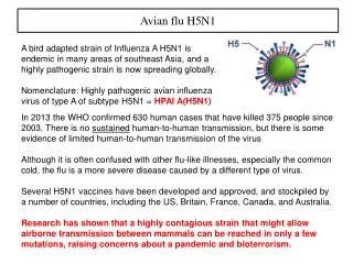
H5N1 The deployment • Evaluate potential targets and model their 3D structures • Prepare the large-scaledocking using Autodock. • Development of the grid environment for a large-scale deployment. From Prof. Ying-Ta Wu et al.
Preparation of neuraminidase 1: GH family 34/H5N1 Ni-NTA column chromatography E.coliRosseta ( DE3 ) haboring-Neu1-23d LB media (5 ml) containing Ampicilline 50 ug/ml Cultured until OD600=0.5 at 37oC Cool down in ice Add IPTG Cultured more …. 16oC For purification, 4 L culture. sp Neu1 : 1350 bp Elution by from 25 to 500 mM IMD Each fraction: 15 ml SDS-PAGE (12%) of Neu1 kDa 209 124 80 Neuraminidase 50kDa 49.1 detect on the UV llumination 34.8 20.6 After 4 h 6 8 10 12 14 8 SM SM T S
Preparation of neuraminidase 2: GH family 33/ C. perfringens Neuraminidase Ni-NTA column chromatography E.coliRosseta ( DE3 ) haboring-CP42 LB media (5 ml) containing Ampicilline 50 ug/ml Cultured until OD600=0.5 at 37oC Cool down in ice Add IPTG Cultured more …. 18oC For purification, 1 L culture. Elution by from 25 to 500 mM IMD Each fraction: 15 ml Expression vector SDS-PAGE (10%) of CP42 kDa 209 124 Neuraminidase 80 49.1 42kDa detect on the UV illumination/ Florescent at excitation at 332 nm emission 448 nm 34.8 20.6 CE SM 8 10 12 14 S CP42
Assay for neuraminidase activity 2: 4-MU-NANA 4-Methylumbeliferyl-N-acetyl-a-D-neuramininic acid ammonium salt [4MU-NANA]; Substrate First screening (200 nmol) Recombinant Neuraminidase Spectrofluorometric detector RF-551 362 nm excitation and 448 nm emission wavelengths Second screening (2 nmol) Red Kinetic study Inhibition Blue
Screening of neuraminidase: First screening Neu1 in GH 34 CP42 in GH 33 First Screening 116/308 compounds – 38% First Screening 42/169 compounds – 25%
Screening of neuraminidase: Second screening Neu1 in GH 34 CP42 in GH 33 Second screening 62/308 compounds-20.1% (Higher inhibition activity compare to Tamiflu) Second screening 0/169 compounds-0%
4MU-NANA : 20 mM/RM Neuraminidase : 10 mU/reaction Measure at excitation 362 nm and emission at 448 nm On UV
Preparation of H5N1 mutant enzymes: GH family 34/N1Mutation Megar primer PCR 1. FPCR 2. Second PCR 3. Cloning 7 mutants E119G/A/D/V H274Y/F R292K N294S From Prof. Ying-Ta Wu et al.
Acknowledgements Enzyme in vitro tests: Chonnam National University, Korea Young-Min KIM (Nuraminidases), Hee-Kyoung KANG (Plasmepsin) Nuraminidase, Plasmepsin In silico data challenge and analyses: Academia Sinica, Taiwan Hurng-Chun LEE, Simon C. LIN (Grid Computing Center) Ying-Ta WU, Chon-Chen LEE (Genomic Research Center) CNRS-IN2P3-LPC, Clermont-Fd, France Vincent BRETON, Nicolas JACQ, Jean SALZEMANN Vinod KASAM, Ana DA COSTA, Vincent BLOCH Yannick LEGRE HealthGrid Nicolas SPALINGER SCAI-Fraunhofer Institute, Germany Martin HOFMANN Modena University, Italy Giulio RASTELLI