Download
1 / 83
920 likes | 1.4k Views
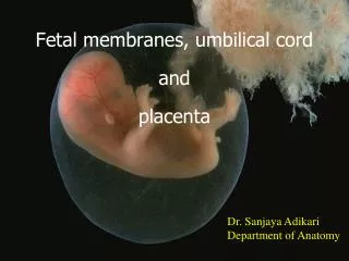
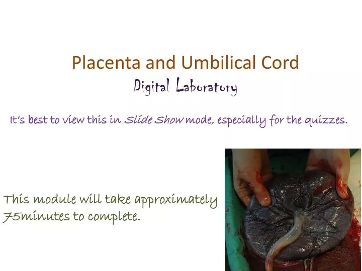
Placenta and Umbilical Cord Digital Laboratory. It’s best to view this in Slide Show mode, especially for the quizzes. This module will take approximately 75minutes to complete. After completing this exercise, you should be able to:

E N D
Placenta and Umbilical Cord Digital Laboratory It’s best to view this in Slide Show mode, especially for the quizzes. This module will take approximately 75minutes to complete.
After completing this exercise, you should be able to: • identify, at the light microscope level, each of the following: • Placenta – note you will not need to distinguish the developmental age of placenta • Fetal portion • Chorion • Amnion • Stem villi • Branch villi • Mesenchyme • Cytotrophoblast • Syncytiotrophoblast • Syncytial knots • Anchoring villi (same substructures as stem villi) • Maternal portion • Basal plate • Fibrinoid • Decidual tissue • Myometrium • Deciduaparietalis • Decidual cells • Umbilical cord • Amnion • Mesenchyme • Umbilical arteries and veins • identify, at the electron microscope level, each of the following: • Placenta • Cytotrophoblasts • Syncytiotrophoblast
EMBRYOLOGY REVIEW Review early embryology by clicking on this audio file.
EMBRYOLOGY REVIEW Review early embryology by clicking on this audio file. Endometrial epithelium
EMBRYOLOGY REVIEW Review early embryology by clicking on this audio file.
EMBRYOLOGY REVIEW Review early embryology by clicking on this audio file. Chorionic cavity = extraembryonic coelom Chorionic cavity Chorionic cavity
EMBRYOLOGY REVIEW Review early embryology by clicking on this audio file. amniotic cavity germdisk amnioblasts/ amniotic membrane extraembryonic mesoderm yolksac
EMBRYOLOGY REVIEW Review early embryology by clicking on this audio file.
GROSS ANATOMY OF THE PLACENTA • The placenta that is ejected has two sides: • a rough maternal side composed of decidual (endometrial) tissue • A smooth fetal side composed of the amniotic membrane Fetal side maternal side maternal side Fetal side
GROSS ANATOMY OF THE PLACENTA • The placenta is formed by the joining of three structures: • Amnion (amniotic membrane) • Chorion (including region containing villi) • Maternal decidua The chorion is tightly attached to the maternal decidua when the conceptus implants into the uterine lining. In contrast, the amnion is only loosely opposed to the chorion, and these layers will separate during tissue preparation.
GROSS ANATOMY OF THE PLACENTA • The placenta is formed by the joining of three structures: • Amnion (amniotic membrane) • Chorion • Maternal decidua Our discussion of the placenta will start on the fetal side with the amnion and chorion, and progress in the direction of the arrow. The first placental slide is oriented opposite this drawing, so the amniotic fluid is to the right, and the maternal tissue is to the left. The images are from a region similar to that within the blue box, and includes the amnion, chorion, and villi.
AMNION AND CHORION chorion The fetal side is composed of the amnion and the chorion. villi chorion amnion Amniotic fluid would be here
AMNION AND CHORION Extraembryonic mesoderm of chorion The green dotted line represents the approximate location of the obliterated chorionic cavity, which is NOT obliterated in this image. Extraembryonic mesoderm of amnion Amniotic fluid would be here • The chorion is composed of three layers: • Extraembryonicmesenchyme • Cytotrophoblasts (yellow arrows) • Syncytiotrophoblasts (red arrows) • Closer examination of the amnion reveals it consists of two layers: • Amniotic epithelium (amnioblasts, blue arrows) • Extraembryonicmesenchyme
AMNION AND CHORION Video showing amnion and chorion in placenta at 5 months – SL145 • Link to SL 145 • Be able to identify: • Amnion • Amnioblasts • Extraembryonicmesenchyme • Chorion • Extraembryonicmesenchyme • Trophoblasts • Cytotrophoblast and syncytiotrophoblasts differentiated on a subsequent slide
AMNION AND CHORION chorion This is a different slide than the previous one, so it looks a little different. The space between the amnion and chorion is the extraembryoniccoelom, or chorionic cavity. This is a potential space in the placenta that is recreated in our tissue sections. Placental villi amnion Chorionic cavity Amniotic fluid would be here
AMNION AND CHORION X Clotted blood X Extraembryonic mesoderm of amnion Amniotic fluid would be here Extraembryonic mesoderm of chorion The chorionic cavity indicated by the Xs. Maternal blood has clotted against the trophoblast cells, making it difficult to identify them in this image. X • Closer examination of the amnion reveals it consists of two layers: • Amniotic epithelium (amnioblasts, blue arrows) • Extraembryonicmesenchyme • The chorion is composed of three layers: • Extraembryonicmesenchyme • Cytotrophoblasts • Syncytiotrophoblasts
AMNION AND CHORION Video showing amnion and chorion in placenta at term – SL146 • Link to SL 146 • Be able to identify: • Amnion • Amnioblasts • Extraembryonicmesenchyme • Chorion • Extraembryonicmesenchyme • Trophoblasts • Cytotrophoblast and syncytiotrophoblasts differentiated on a subsequent slide
VILLI AND INTERVILLOUS SPACE chorion Villi that project directly from the chorion are called stem villi. You can see numberous branches from the stem villi, called branch villi. The empty space between the villi is normally filled with maternal blood, and is called the intervillous space. villi stem villus amnion Amniotic fluid would be here
VILLI AND INTERVILLOUS SPACE chorion In the placenta at term, note the number of villi has increased dramatically, providing more surface area that increases the efficiency of nutrient and waste exchange, supporting the increasing demands of the growing fetus. villi amnion Amniotic fluid would be here
VILLI AND INTERVILLOUS SPACE The next images are taken from within the villous space (blue box).
VILLI AND INTERVILLOUS SPACE villus This region shows villi and the intervillous space. Remember the intervillous space develops from the trophoblastic lacunae that formed within the syncytiotrophoblast. villus The next slide is an enlarged region in the blue box. villus
VILLI AND INTERVILLOUS SPACE syncytiotrophoblast • Like the chorion, villi contain: • mesenchyme with blood vessels • cytotrophoblasts – large, euchromatic nuclei with pale cytoplasm • syncytiotrophoblasts – clustered nuclei with darker cytoplasm cytotrophoblast blood vessels mesenchyme We’ll see better cytotrophoblasts on the next slide.
VILLI AND INTERVILLOUS SPACE syncytiotrophoblast • In this magnified image: • mesenchyme with blood vessels • cytotrophoblasts – large, euchromatic nuclei with pale cytoplasm • syncytiotrophoblasts – clustered nuclei with darker cytoplasm cytotrophoblasts Intervillous space (with mama’s blood) Red blood cell in blood vessel cytotrophoblasts mesenchyme
VILLI AND INTERVILLOUS SPACE Video showing villi – SL145 • Link to SL 145 • Be able to identify: • Villi • Stem villi • Branch villi • Mesenchyme • Cytotrophoblast • Syncytiotrophoblast • Intervillous space • Where is fetal blood? Where is maternal blood?
VILLI AND INTERVILLOUS SPACE • As the placenta matures, changes occur to increase exchange efficiency: • Villi branch extensively • Cytotrophoblasts decrease in number • Syncytiotrophoblast nuclei cluster, forming syncytial knots (yellow circles) - this allows the remainder of the syncytiotrophoblast to thin • Fetal blood vessels move to the edge of the villi, where the basal lamina of the endothelial cells fuses with the trophoblast basement membrane
VILLI AND INTERVILLOUS SPACE Better images of syncytial knots (yellow circles) – the one in the right image is sectioned so it appear to be floating within the intervillous space.
VILLI AND INTERVILLOUS SPACE Video showing villi – SL146 • Link to SL 146 • Be able to identify: • Same as previous slide, including • Increased number of villi • Lack of cytotrophoblasts • Syncytial knots • Areas of fused basal lamina
VILLI AND INTERVILLOUS SPACE This electron micrograph focuses on the barrier between maternal blood (ME) and fetal blood (FE) at term. The syncytiotrophoblast (syn) is thin, with multiple nuclei (only one, N, shown here). This mass is very active metabolically, with microvilli, rough and smooth ER, Golgi, secretory vesicles, and lipid droplets. There is no cytotrophoblast in this section. The basement membrane of the trophoblast (TBL) and the endothelial cell of the fetal blood vessel (EBL) are separated by a thin layer of connective tissue here.
MATERNAL SIDE OF PLACENTA The next images are taken from the maternal side of the placenta (blue box).
MATERNAL SIDE OF PLACENTA • The maternal side of the placenta shows: • Villi • Decidua • Myometrium myometrium decidua villi The next slide shows an image taken from a region similar to the one within the yellow rectangle.
MATERNAL SIDE OF PLACENTA Some villi extend across the intervillous space and make contact with the syncytiotrophoblasts that line the decidual tissue. These anchoring villihave many cytotrophoblasts (e.g. many within red dashed line), which are moving between the syncytiotrophoblasts and decidual tissue to form the cytotrophoblastic shell. Maternal decidual cells and blood form highly eosinophilicfibroid. The cytotrophoblastic shell is not readily apparent on our slides. Anchoring villus The syncytiotrophoblasts, cytotrophoblastic shell, and maternal decidual tissue is collectively called the basal plate.
MATERNAL SIDE OF PLACENTA decidua villi • In this image, review anchoring villiwith cytotrophoblasts, and fibrinoid. • In the decidua, you can see many large cells with eosinophilic cytoplasm. These cells are: • Decidual cells • Cytotrophoblasts • Syncytiotrophoblast • It is difficult to distinguish these three on our slides, though multinuclear masses are clearly syncytiotrophoblast.
MATERNAL SIDE OF PLACENTA Video showing maternal side of placenta – SL145 • Link to SL 145 • Be able to identify: • Villi • Anchoring villi • cytotrophoblasts • Decidua • Fibrinoid • Large, eosinophilic cells (decidual or trophoblastic cells) • Myometrium
MATERNAL SIDE OF PLACENTA At term, there is substantial fibrinoid in the decidua, with many eosinophilic cells.
VILLI AND INTERVILLOUS SPACE Video showing maternal side of placenta – SL146 • Link to SL 146 • Be able to identify: • Villi • Anchoring villi • cytotrophoblasts • Decidua • Fibrinoid • Large, eosinophilic cells (decidual or trophoblastic cells) • Myometrium
DECIDUA PARIETALIS The next images are taken from the decidualparietalis (blue box).
DECIDUA PARIETALIS In this low power image, you can see the myometrium and decidua. myometrium decidua Note the numerous endometrial glands. The next slide is an image similar to the region in the yellow rectangle.
DECIDUA PARIETALIS • In this image, you can see • Thin endometrial epithelium (green arrows) • Extensive vasculature (red arrows) • Decidual cells (blue arrows) – large, with eosinophilic cytoplasm. Unlike the previous slide, these must be decidual cells because they are in the deciduaparietalis.
DECIDUA PARIETALIS Video showing decidua parietalis – SL147 • Link to SL 147 • Be able to identify: • Deciduaparietalis • Endometrial glands • Extensive vasculature • Decidual cells
DECIDUA PARIETALIS At birth, the placenta and the remainder of the functional region of the endometrium slough off, leaving the basal region behind to regenerate the endometrium (similar to the normal menstrual cycle). In this image of the decidua parietalis, you can see that epithelial cells of the functional zone have atrophied (black bracket), while those in the basal region remain columnar. myometrium
UMBILICAL CORD • The left image is a scanning view of the umbilical cord at 5 months. The image to the right is an enlargement of a region similar to that in the blue box. Note: • The outer layer of the umbilical cord is epithelial (amnioblasts) • The core of the umbilical cord is mesenchyme • There is a central umbilical vein, flanked by two umbilical arteries
UMBILICAL CORD Video showing umbilical cord at 5 months – SL35 • Link to SL 035 • Be able to identify: • Umbilical cord • Amnioblasts • Mesenchyme • Umbilical vein • Umbilical arteries
UMBILICAL CORD Umbilical cord at term. Note that the mesenchyme is more fibrous and the blood vessels are more developed.
UMBILICAL CORD Video showing umbilical cord at term – SL148 • Link to SL 148 • Be able to identify: • Umbilical cord • Amnioblasts • Mesenchyme • Umbilical vein • Umbilical arteries
The next set of slides is a quiz for this module. You should review the structures covered in this module, and try to visualize each of these in light micrographs: • identify, at the light microscope level, each of the following: • Placenta – note you will not need to distinguish the developmental age of placenta • Fetal portion • Chorion • Amnion • Stem villi • Branch villi • Mesenchyme • Cytotrophoblast • Syncytiotrophoblast • Syncytial knots • Anchoring villi (same substructures as stem villi) • Maternal portion • Basal plate • Fibrinoid • Decidual tissue • Myometrium • Decidua parietalis • Decidual cells • Umbilical cord • Amnion • Mesenchyme • Umbilical arteries and veins • identify, at the electron microscope level, each of the following: • Placenta • Cytotrophoblasts • Syncytiotrophoblast
Final quiz Self-check: Identify the organ. (advance slides for answers) Uterus, secretory phase
QUIZ Self-check: Identify the region indicated by the brackets. (advance slides for answers) Adrenal medulla
Final quiz Self-check: Identify the cells indicated by the arrows. (advance slides for answers) syncytiotrophoblast
QUIZ Self-check: Identify the tissue closest to the arrows (advance slides for answers) Transitional epithelium
Final quiz Self-check: Identify the outlined organ. (advance slides for answers) Posterior pituitary