Download
1 / 45
530 likes | 906 Views
Approach to Anemia/ PM. Abdallah Abbadi. MD.FRCP Professor of Medicine, Hematology & Oncology Jordan University & University Hospital Email: aabbadi@ju.edu.jo aawidi@yahoo.com. Anemia. Defined as low Hb for that age and gender Understanding anemia

E N D
Approach to Anemia/ PM Abdallah Abbadi. MD.FRCP Professor of Medicine, Hematology & Oncology Jordan University & University Hospital Email: aabbadi@ju.edu.jo aawidi@yahoo.com
Anemia • Defined as low Hb for that age and gender • Understanding anemia • Disease - to be treated on its own merits • Condition - a secondary manifestation of another disease • Causes • Decreased production • Blood loss • Hemolysis
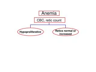
Classification of Anemia • Clinical findings • Acute • Chronic • Red cell kinetics • Determined by reticulocyte count • Red cell size • Determined by MCV
General Formulas in anemia Retics Production Index(RPI) = retics x hct÷nhct/2 (N 1-2) MCV= hct x 10/RBC in mill (N 82-92) MCH= Hb g/dl x 10 /RBC in mill (N 27-32) MCHC= Hb g/dl x 100 /hct% (N 32-36) RDW = (Standard deviation of red cell volume ÷ mean cell volume) × 100
Anemia? Production? Survival/Destruction? Bleeding? The key test is the Retics count …..
The reticulocyte count(kinetic approach) • Increased reticulocytes (greater than 2-3% or 100,000/mm3 total) are seen in blood loss and hemolytic processes, although up to 25% of hemolytic anemias will present with a normal reticulocyte count due to immune destruction of red cell precursors. • Retic counts are most helpful if extremely low (<0.1%) or greater than 3% (100,000/mm3 total).
The reticulocyte count • To be useful the reticulocyte count must be adjusted for the patient's hematocrit. Also when the hematocrit is lower reticulocytes are released earlier from the marrow so one can adjust for this phenomenon. Thus: • Corrected retic. = Patients retic. x (Patients Hct/45) • Reticulocyte index (RPI) = corrected retic. count/Maturation time (Maturation time = 1 for Hct=45%, 1.5 for 35%, 2 for 25%, and 2.5 for 15%.) • Absolute reticulocyte count = retic x RBC number.
A 43 yo man is brought to the OPD for evaluation. He was reported to be unwell for 2 months. He is has lost weight and looks cachectic. He was found to have cervical lymphadenopathy. PMH is significant for multiple traumas. Labs Hgb 10 Hct 30 MCV 88 wbc 4.1 plts 120,000 Bun 42 Cr 1.2 Retic. Ct. 1.1% Hemoccult negative
Causes of Anemia (kinetic approach) • Decreased erythrocyte production • Decreased erythropoietin production • Inadequate marrow response to erythropoietin • Erythrocyte loss • Hemorrhage • Hemolysis
Anemia: Etiologies • Production defects: Nutritional deficiencies - Vitamin B12, folate or iron deficiency. Inflammation/chronic disease. Primary marrow disorders- pure red cell aplasia, myelodysplasia. • Sequestration(hypersplenism)-usually associated with mild pancytopenia. • Blood loss. • Blood destruction.
MCV>115 B12, Folate Drugs that impair DNA synthesis (AZT, chemo., azathioprine) MDS MCV 100 - 115 Ditto endocrinopathy (hypothyroidism) reticulocytosis Underproduction (1)(morphological approach)
Normocytic Anemia of chronic disease Mixed deficiencies Renal failure Microcytic Iron deficiency Thal. trait Anemia of chronic disease (30-40%) sideroblastic anemias Underproduction (2)
A 29 yr female artist is referred to you because of anemia which has not responded to oral iron therapy. PMH: She had gastric partition 3 yrs ago and has lost a lot of weight. PE shows a pale woman, liver and spleen are not enlarged, stool guaiac is negative. Labs Hgb 8.8 g/dl, MCV 75 fl WBC 5500/ul Plts 490,000/ul, Retic ct 20,000/ul Fe 25 ug/dl, TIBC 460 ug/dl, ferritin 11 ug/l
Iron deficiency is a common form of malnutrition that affects more than 2 billion people globally.
Esophageal webs and strictures Koilonychia Behavioral and neuropsychiatric manifestations Pica (pagophagia) Angular stomatitis Glossitis Systemic Manifestations of Iron Deficiency
Prevalence (%) of iron deficiency and iron-deficiency anemia, United States, third National Health and Nutrition Examination Survey, 1988–199. Sex and age (years)Iron deficiency Iron-deficiency anemia Both sexes 1–2 9 3* 3–5 3 <1 6–11 2 <1 Nonpregnant females 12–15 9 2* 16–19 11* 3* 20–49 11 5* 50–69 5 2 ³ 70 7* 2* *Prevalence in non-blacks is 1 percentage point lower than prevalence in all races.
Inadequate iron supply • Poor nutritional intake in children (not a common independent mechanism in adults but often a contributing factor) • Malabsorption • Gastric bypass surgery for ulcers or obesity • Achlorhydria from gastritis or drug therapy • Severe malabsorption (for example, celiac disease [nontropical sprue]) • Abnormal transferrin function • Congenital atransferrinemia • Autoantibodies to transferrin receptors
Oral iron failure? • Incorrect diagnosis (eg, thalassemia) • anemia of chronic disease? • Patient is not taking the medication • Not absorbed (enteric coated?) • Rapid iron loss?
Intravenous Iron Therapy • 60 kg woman with a hgb of 8: • Her total blood volume should be 3900 mL or 39 deciliters (65 mL/kg x 60 kg). • A normal hemoglobin concentration would be 14 g/dL. Thus, her hemoglobin deficit is 6 g/dL with a total deficit of 234 g (6 g/dL x 39 dL). • Each gram of hemoglobin contains 3.3 mg of iron. Thus, her total red cell iron deficit is 772 mg (234 g of hemoglobin x 3.3 mg Fe per gram). For iron dextran/ sucrose: 0.5 mL test dose is given IV over at least 30 seconds, remainder given at a rate not exceeding 50 mg (one mL) per minute, and a total dose not exceeding 100 mg (two mL) per day
55 yr F with moderately severe Rheumatoid Arthritis taking Prednisone 10 mg/day, Celecoxib, is referred to you for an anemia workup • CBC: Hct = 30%, MCV = 82, WBC = 5.4 thou/l, plt = 345 thou/ l • Smear - Normal • Retic count = 2 % (Corrected Retic = 30/40 x 2%= 1.5%) • Fe = 20 g/dL (55-155), TIBC = 200 g/dL (270-400), Transferrin saturation = 20/200 = 10% (15-50) • Ferritin = 330 g/dL (20-160)
Iron Deficiency Anemia vs. Inflammatory Block • Smear: • hypochromic and microcytic (low MCV) RBCs, usually not seen unless Hct 30% • platelet count is often elevated • Ferritin: a measure of total body iron stores, but also an acute phase reactant • <15g/l = Fe deficiency, 150 g/l = Not Fe deficiency15-150 g/l = ?
Iron Deficiency Anemia vs. Inflammatory Block • Low Iron Saturation (Fe/TIBC ratio) • Fe (not reliable) • TIBC • Fe/TIBC (% saturation) 15% • BM bx: absent Fe stores • Gold standard • Therapeutic Trial of Oral Iron
Utility of supraphysiologic doses of erythropoietin in the setting of inflammatory block.
Fe-deficiency Rheumatoid arthritis Baer AN, et al. Blunted erythropoietin response to anemia in rheumatoid arthritis. Br J Haematol. 1987;66:559–64.
A 69 yo woman is referred to you for progressive anemia. The most recent blood counts reveal leukopenia and thrombocytopenia. Examination of the peripheral blood shows hypersegmented granulocytes. The neurologic examination is normal, and her serum folate is normal. CBC: Hb 9, MCV 105, RDW 15, Retics (corrected <0.1%), WBC 3500 (n diff), Plt 103. DAT is –ve.
B12/Folate Deficiency (2) • Dx: • Smear: Macrocytic (High MCV) RBCs, +/- hypersegmented neutrophils, +/- modest neutropenia, but… • the diagnosis of B12 def. was made in patients in whom only 29 percent had anemia, and only 36 percent had a MCV greater than 100 fL (Pruthi RK, Tefferi A, Mayo Clin Proc 1994 Feb;69(2):144-50) • B12 • Low serum B12, elevated serum methylmalonic acid levels • Anti-IF Abs, Schilling test (?), PA accounts for 75% • Folate • Serum folate level-- can normalize with a single good meal
B12/Folate Deficiency (3) • Tx: • B12 deficiency: B12 1 mg/month IM, or 1-2 mg/day po • Folate deficiency: Improved diet, folate 1 mg/day • Monitor for a response to therapy. • Pernicious Anemia – monitor for gi cancers.
Cobalamin deficiency and neurological problems • Subacute combined degeneration of the dorsal and lateral spinal columns. • Well known study of B12 deficiency in the nursing home population • Vitamin B-12 deficiency is present in up to 15% of the elderly population • Is oral B12 good enough? • Association between nitrous oxide anesthesia and development of neurological symptoms responsive to B12 in patients with subclinical cobalamin deficiency
Sideroblastic Anemias • Heterogenous grouping of anemias defined by presence of ringed sideroblasts in the BM • Etiologies: • Hereditary (rare), type of porphyria • Myelodysplasia • Tx: • Trial of pyridoxine for hereditary or INH induced SA
A 32-year-old woman has Crohn’s disease that has waxed and waned for 15 years. A recent flare beginning 2 weeks ago was treated with sulfasalazine and corticosteroids. Despite improvement in diarrhea and abdominal pain, she continues to feel ill and experiences easy fatigability with dyspnea and palpitations on mild exertion. On physical examination, pallor, trace scleral icterus, and active bowel sounds are noted. Laboratory studies show: hematocrit, 22%; leukocyte count, 14,000/ul. (90% polymorphonuclear neutrophils with shift to the left); reticulocyte count, 7%; platelets noted to be “adequate on smear.”
Hemolytic Anemias Hemolytic anemias are either acquired or congenital. The laboratory signs of hemolytic anemias include: 1. Increased LDH (LDH1) - sensitive but not specific. 2. Increased indirect bilirubin - sensitive but not specific. 3. Increased reticulocyte count - specific but not sensitive 4. Decreased haptoglobin - specific but not sensitive. 5. Urine hemosiderin - specific but not sensitive.
General Principles • Anemia is a sign, not a disease. • Anemias are a dynamic process. • Its never normal to be anemic. • The diagnosis of iron deficiency anemia mandates further work-up.
A GP has referred a 21-year-old married woman for evaluation of her recently documented anemia. HPI: She was recently married and wants to have a family, but went to her GP because she felt that she had less energy than her friends. She has no history of melena or bright red blood per rectum and her menstrual history seemed normal. She thinks that her mother and 2 maternal aunts have anemia. Physical examination: She is a pale but otherwise alert, healthy young woman. No scleral icterus is present and her chest and heart exam are normal. A soft spleen tip is palpable in the left upper quadrant (LUQ). No edema is present. Labs: White blood cells (WBC) 4600, normal differential, platelets 421,000/ul, hematocrit (Hct) 27, hemoglobin (Hgb) 8.1gm/dl, red blood cells (RBC) 4.58M/ul, MCV 59, mean corpuscular hemoglobin (MCH) 17, mean corpuscular hemoglobin concentration (MCHC) 30. Retic 3.1% Absolute retics 142,000/ul, ferritin 482 ng/ml, serum iron 149, transferrin 193, % sat 77%.
The hemoglobin electrophoresis reveals HbA2 is 1%, HbF is 0.5%, and HbH is 16%.
Thalassemias • Genetic defect in hemoglobin synthesis • synthesis of one of the 2 globin chains ( or ) • Imbalance of globin chain synthesis leads to depression of hemoglobin production and precipitation of excess globin (toxic) • “Ineffective erythropoiesis” • Ranges in severity from asymptomatic to incompatible with life (hydrops fetalis) • Found in people of African, Asian, and Mediterranean heritage
Thalassemias (2) • Dx: • Smear: microcytic/hypochromic, misshapen RBCs • -thal will have an abnormal Hgb electrophoresis (HbA2,HbF) • The more severe -thal syndromes can have HbH inclusions in RBCs • Fe stores are usually elevated • Tx: • Mild: None • Severe: RBC transfusions + Fe chelation, Stem cell transplants