Download

1 / 38
420 likes | 1.11k Views
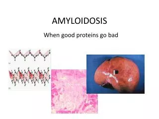
Amyloidosis. Amyloid is a class of insoluble, homogeneous, eosinophilic ("hyaline") substances that accumulate in extracellular spaces. Their common link is that they are beta-pleated sheets (rather than α helices), and therefore cannot be effectively handled by the body.

E N D
Amyloid is a class of insoluble, homogeneous, eosinophilic ("hyaline") substances that accumulate in extracellular spaces. Their common link is that they are beta-pleated sheets (rather than α helices), and therefore cannot be effectively handled by the body. • All amyloids are now known to be built up from soluble oligomers with a particular conformation that seems necessary to their becoming beta-pleated. • Amyloids don't go away, and eventually the surrounding cells undergo "pressure atrophy". • Whatever is really happening, enough amyloid in an organ makes it rubbery and waxy and (sometimes) useless. Amyloidosis is a group of curious systemic and localised diseases with varied and bizarre clinical presentations. • The classic forms of amyloidosis are not among the commonest diseases, though they are not rare. • Alzheimer's disease, now recognized to be one of the commonest and deadliest diseases, regularly includes amyloid beta/A4 deposition in the brain and its vessels. • Amyloidosis involving the heart is responsible for an unknown number of cases of heart disease in the elderly.
Most amyloids are altered forms of various proteins that exist in the healthy body. • Regardless of its origin and amino-acid sequence, amyloid is not an alpha-helix, but a crossed beta-pleated sheet. • This beta-pleated structure results in the marked affinity all amyloids exhibit for Congo Red dye, and for the apple-green birefringence it exhibits when so stained and examined under polarized light. • Congo red: • amyloid stains metachromatically (i.e., a different color from the dye solution, in this case red rather than violet) with crystal violet • has other exotic reactions with special stains (notably fluorescence with thioflavin T).
Despite its amorphous appearance on light microscopy, electron microscopy shows that 90% of any type of amyloid consists of non-branching fibrils, 70 to 100 angstroms across. These are usually crisscross but may be parallel. • These fibrils are the beta-pleated protein, the congophilic component of amyloid. • The remaining 10% of any amyloid is composed of "P-component", a 90-angstrom, pentagonal ‘doughnut-rod thing’. • The P component is pentamers of a glycosylated α-1 globulin (serum amyloid P-component - SAP), an acute-phase protein similar in sequence to C-reactive protein that is produced by the liver in response to interleukin 1 production by macrophages. It imparts the weak PAS-positivity to amyloid. • The P component, bound to the fibrils, is also required to prevent the amyloid from being degraded by the body. • As noted, all types of amyloid look the same at any magnification.
This Congo red stain reveals orange-red deposits of amyloid Amyloidosis
This is the immunofluorescent appearance of the myocardium with antibody to lambda light chain. Thus, this is "AL amyloid". Amyloidosis
By electron microscopy, amyloid is seen to be composed of a "beta-pleated sheet" of fibrils, seen here as irregular grey material. When the amyloid protein is made up of immunoglobulin light chains, then it is "AL amyloid" and when it is derived from serum amyloid-associated protein, then it is "AA amyloid." Amyloidosis
Deposits expand mesangium and encroach on glomerular capillary lumen. Filaments are haphazardly arranged, hollow, non-branching and about 10 nm in diameter, especially perpendicular to the basement membrane Amyloidosis
Red Arrow – atropic hepatocytesBlack Arrow – amyloid deposition
When stained with Congo red and observed under polarized light, the amyloid has a characteristic "apple green" birefringence as seen here in a deposit around an artery in the heart. Amyloidosis Heart
AmyloidosisHeart In any form of systemic amyloidosis, involvement of the heart may be severe or mild. Amyloid deposition begins in the subendocardium. In relatively mild cases, tiny deposits of amyloid occur on the atrial endocardium that resemble dewdrops. But more often, there's nothing at all abnormal grossly. Almost all elderly people have traces of amyloids of unknown composition in their aortas, independent of atherosclerosis, as well as deposits of atrial natriuretic peptide (amyloid IAA). These are probably harmless. Of course, many will also have some transthyretin-based amyloid, and most of these people are probably asymptomatic. The heart in severe cardiac amyloidosis, however, is stiff and heavy. This is the usual cause of "restrictive cardiomyopathy" (stiff-heart disease).
AmyloidosisLardaceous Spleen Amyloid deposition in the spleen is usual in systemic amyloidosis. Most patients get increased circulating platelets, but the relative loss of splenic function is among the least of these peoples' problems. There are two classic, unappetizing descriptions of amyloid-loaded spleens seen at autopsy. A "sago spleen" has its amyloid in the white pulp. A "lardaceous spleen" has its amyloid in the red pulp (and usually indicates amyloid B). It looks like lard.
Here is a chronic renal disease that may actually increase the size of the kidney. Pale deposits of amyloid are present in the cortex, most prominently at the upper center. Amyloid gets deposited in the glomeruli, the blood vessels, and around the tubules. Usually the glomerular basement membrane becomes too leaky to proteins, and the patient gets the "nephrotic syndrome". If the patient with renal amyloidosis survives, eventually the amyloid plugs up the glomeruli and that's the end of the kidneys. Renal involvement is a major cause of death in amyloidosis. Amyloidosis Kidney
This is dystrophic calcification in the wall of the stomach. At the far left is an artery with calcification in its wall. There are also irregular bluish-purple deposits of calcium in the submucosa. Calcium is more likely to be deposited in tissues that are damaged. Calcification, dystrophic Stomach
Microscopically, the infiltrating ductal carcinoma extends irregularly through the tissue as cords and nests of neoplastic cells with intervening collagen. There is a purplish microcalcification at the lower center right. Neoplastic cells are not as robust or as organized as normal cells and are more likely to undergo necrosis. Dystrophic calcification can occur in these areas. Calcification, dystrophic Breast
There is a severe degree of narrowing in this coronary artery. It is "complex" in that there is a large area of calcification on the lower right, which appears bluish on this H&E stain. Complex atheroma have calcification, thrombosis, or hemorrhage. Such calcification would make coronary angioplasty difficult. Calcification, dystrophic Atherosclerosis
Here is so-called "metastatic calcification" in the lung of a patient with a very high serum calcium level (hypercalcaemia). Calcification, metastatic Lung
The black streaks seen between lobules of lung beneath the pleural surface are due to accumulation of anthracotic pigment. This anthracosis of the lung is not harmful and comes from the carbonaceous material breathed in from dirty air typical of industrialized regions of the planet. Persons who smoke would have even more of this pigment. Anthracosis Lung
By polarized light microscopy can be seen the aetiology for most pneumoconioses (even those in coal miners) silica crystals. Here are seen bright white crystals of varying sizes. The silica induces a fibrogenic response by macrophages to produce the nodular foci of collagen deposition. Pneumoconioses Lung
When silica dust is inhaled into the lungs, the tiny particles are engulfed by macrophages, large phagocytic cells that play a key role in fighting off bacteria and other foreign substances. The cells are unable to digest the dust and the silica kills them. The dead macrophages slowly accumulate in the lungs so that they eventually form fibrous nodules, which may further coalesce into sizable masses. These fibrous masses, which are characteristic of silicosis, hinder lung expansion and gas exchange. Thus, the primary symptoms of silicosis are breathing difficulty, coughing, chest pain, and general weakness. Silicosis is a disease of the lungs caused by excessive or chronic exposure to silica dust. Silica is the chief mineral component of sand and is found in many varieties of rock and in mineral ores. Thus, individuals employed in certain occupations, such as sandblasting, mining, grinding, and drilling, are at increased risk for developing the disease. Silicosis
Studies indicate that many people with tattoos wish they had never received them. Unexpected break-ups, changes in lifestyles, and similar reasons are frequently to blame. Yet, for those who can afford it, tattoos can often be removed. Laser treatments that break the pigment molecules comprising a tattoo into smaller particles that can be eradicated by the cells of the immune system and are associated with only a small risk of permanent scarring are generally the preferred method of tattoo removal. Other treatments, including dermabrasion, surgical excision, and cryosurgery, are also sometimes employed, but are typically more painful and likely to cause scarring than the laser technique. Tattoos are created by injecting or otherwise ingraining colored pigments into the dermal layer of the skin. Unlike the epidermis, which is continually sloughed by the body, the dermis stays intact throughout one’s lifetime, which makes tattoos permanent. Tattoo
The brown coarsely granular material in macrophages in this alveolus is haemosiderin that has accumulated as a result of the breakdown of RBC's and release of the iron (in haeme). The macrophages clear up this debris, which is eventually recycled. Haemosiderin Deposition Alveolar
These renal tubules contain large amounts of hemosiderin, as demonstrated by the Prussian blue iron stain. This patient had chronic haematuria. Haemosiderin Deposition Renal Tubules
The dark brown color of the liver, as well as the pancreas (bottom center) and lymph nodes (bottom right) on sectioning is due to extensive iron deposition in a middle-aged man with hereditary hemochromatosis (HHC). HHC results from a mutation involving the hemochromatosis gene (HFE) that leads to increased iron absorption from the gut. The prevalence is between 1:200 and 1:500 persons. About 1 in 10 persons of northern European ancestry carries the abnormal recessive HFE gene. Haemochromatosis Liver – Post Motem
A Prussian blue iron stain demonstrates the blue granules of hemosiderin in hepatocytes and Kupffer cells. Hemochromatosis can be primary (the cause is probably an autosomal recessive genetic disease) or secondary (excess iron intake or absorption, liver disease, or numerous transfusions). Hemochromatosis leads to bronze pigmentation of skin, diabetes mellitus (from pancreatic involvement), and cardiac arrhythmias (from myocardial involvement). Haemochromatosis Liver – Prussian Blue Stain
Brown, granular pigments seen here indicate the presence of iron in hepatocytes. Most of the hepatocytes appear pale due to autolysis. Haemochromatosis
Here, the same section has been treated with trichome, which stains areas of fibrosis (blue). Haemochromatosis
The yellow-brown granular pigment seen in the hepatocytes here is lipochrome (lipofuscin) which accumulates over time in cells (particularly liver and heart) as a result of "wear and tear" with aging. It is of no major consequence, but illustrates the end result of the process of autophagocytosis, in which intracellular debris is sequestered and turned into these residual bodies of lipochrome within the cell cytoplasm. Lipofuscin Hepatocytes
Easily seen at the poles of the cardiac nuclei, brown-red in color. The faster the basal rate of metabolism, the faster it produces lipofuscin.Lipofuscin is also the pigment in earwax, giving it a characterisitc yellow-brown color. As one ages, the amount of lipofuscin will continue to increase, especially in the heart. One can determine whether a heart is relatively young or old, as a direct reflection of the amount of lipofuscin present. Lipofuscin Cardiac Cells
Increased amounts of circulating bilirubin in the blood can lead to the physical examination finding of "icterus" or jaundice as seen here from the yellowish hue of the skin. The easiest place to see icterus is on the sclera of the eye. Jaundice
The sclera of the eye is yellow because the patient has jaundice, or icterus. The normally white sclerae of the eyes is a good place on physical examination to look for icterus. Jaundice Scleral icterus
The yellow-green globular material seen in small bile ductules in the liver here is bilirubin pigment. This is hepatic cholestasis. Hepatic Cholestasis