Download
1 / 79

E N D
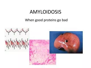
Amyloidosis Definition : In medicine, amyloidosis refers to a variety of conditions in which amyloidproteins are abnormally deposited in organs causing disease. A protein is amyloid if, due to an alteration in its secondary structure, it takes on a particular insoluble form, called the beta-pleated sheet.
AMYLOIDOSIS • Disease characterized by extracellular deposition of pathologic insoluble fibrillar proteins in organs and tissues. • Term amyloid first coined by Virchow in mid 19th century (meaning starch or cellulose). • Amyloid found to stain with congo red, appearing red microscopically in normal light but apple green when viewed in polarized light. • Fibrillar nature and beta pleated sheet configuration described by electron microscopy in 1959.
Misfolded proteins are normally detected and cleared from cell (or stored in aggresomes)
Systemicamyloidoses are those which affect more than one body organ or system. Localisedamyloidoses affect only one body organ or tissue type. Primaryamyloidoses arise from a disease with disordered immune cell function such as multiple myeloma and other immunocytedyscrasias. Secondary (reactive) amyloidoses are those occurring as a complication of some other chronic inflammatory or tissue destructive disease.
Imaging techniques – Technetium Tc 99m pyrophosphate binds avidly to many types of amyloid. Quantitative assessment not possible and strongly positive results usually only occur in pt’s with severe disease. Technetium labeled aprotinin may be more sensitive. • Quantitative scintigraphy can be done with iodine-123- labeled serum amyloid P component (sensitive for AL, ATTR and AA amyloid).
IgH chain translocations are also found in AL especially t(11;14). • Monosomy 18 is also frequently found as well as deletions of chromosome 13. • κ to λ ratio is approx 1:3 • LCD – Non amyloidIg deposition predominantly of κ and usually the constant region. Forms granular rather than fibrillar deposits and mainly affects the kidneys.
Primary Amyloidosis: Conventional Therapy • General measures • Delay target organ failure • Improve quality of life • Specific interventions • Melphalan and Prednisone
Experimental approaches for the treatment of Multiple Myeloma • Allogeneic transplantation (8 studies) • Complete response rate 26-51% • Median event free survival 12-36 months • Revimid (CC-5013) Thalidomide derivative • Phase II study • PS-341 Proteosome inhibitor - Cytotoxic to plasma cells • Phase II study
Feature % Proteinuria/renal insufficiency 91 Diarrhea/malabsorption 22 Goiter 9 Neuropathy/carpal tunnel syndrome 3 Hepatomegaly 5 Lymphadenopathy 2 Cardiac 1 - 2 Presenting Clinical Features in AA Amyloidosis
Macroglossia – occurs in 10-20 % • Amyloid can be found within any part of the GI tact and may infiltrate parenchyma, organs and nerves. • Peripheral neuropathy may be presenting manifestation or develop subsequently during the course of the illness (history of carpal tunnel frequently elicited). • Neuropathy usually distal, symmetric and progressive. Cranial nerve and autonomic nerve involvement also well described. • Motor neuropathy rare.
HEPATIC/SPLENIC • Involvement of liver common. • Hepatomegaly may be striking at presentation and usually disproportionate to extent of liver enzyme abnormalities (except alkaline phosphatase which is frequently elevated). • Presence of jaundice is an adverse prognostic factor and MST from onset of jaundice is only 3 months. • Patients may present with severe intrahepaticcholestasis. • Massive splenic deposition may result in functional hyposplenism.
Plasma-cell-Type Castleman’sdisease with IL-6 release and increased SAA synthesis Amyloid in the lymph node, green birefringence in polarized light Plasma-cell-type Castleman’sdisease (H & E)
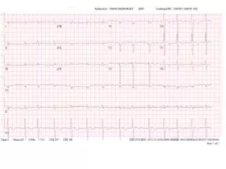
CARDIAC • May present with rapid and progressive onset of CHF. • Characteristically, features are predominantly of right sided CHF. • ECG – low voltage and may have a pattern of MI in absence of CAD. • ECHO – concentrically thickened ventricles with normal-small cavity and diastolic dysfunction on doppler. • Clinical clue is marked worsening of failure when CCB used.
Echocardiogram revealing thickened walls with small chambers
RENAL • Nephrotic syndrome present in 30-50% at diagnosis. • Nephrotic syndrome and renal failure develop only rarely during course of the illness if not present at time of diagnosis. • λ BJP have been associated with inferior survival as compared with κBJP or no monoclonal protein, irrespective of serum creatinine.
Lambda Kappa