Download
1 / 43
430 likes | 446 Views
Learn about the processes of primary and secondary growth in plants, as well as the organization of tissues in stems, leaves, and roots. Discover how cell division and differentiation contribute to the development of the plant body.

E N D
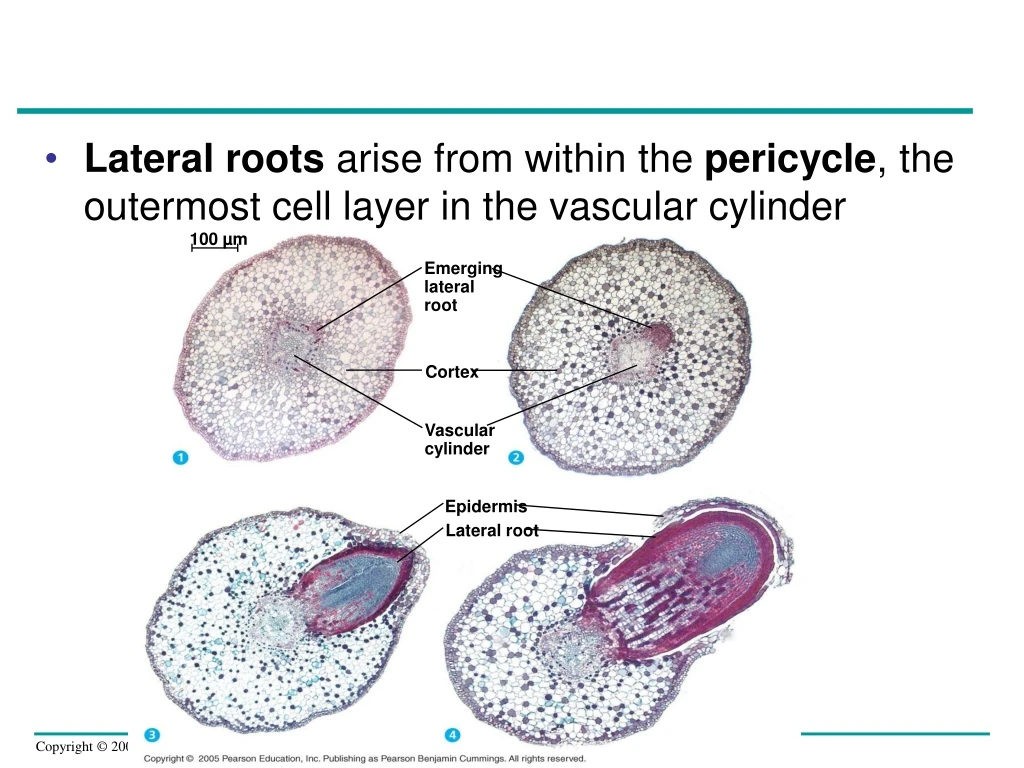
100 µm Emerging lateral root • Lateral roots arise from within the pericycle, the outermost cell layer in the vascular cylinder Cortex Vascular cylinder Epidermis Lateral root
Primary Growth of Shoots Apical meristem Leaf primordia • A shoot apical meristem is a dome-shaped mass of dividing cells at the tip of the terminal bud Developing vascular strand Axillary bud meristems 0.25 mm
LE 35-16 Arrangement of Vascular Tissue Phloem Xylem Ground tissue Sclerenchyma (fiber cells) Ground tissue connecting pith to cortex Pith Epidermis Key Vascular bundles Cortex Epidermis Dermal Vascular bundles Ground Vascular 1 mm 1 mm A monocot (maize) stem. Vascular bundles are scattered throughout the ground tissue. In such an arrangement, ground tissue is not partitioned into pith and cortex. (LM of transverse section) A eudicot (sunflower) stem. Vascular bundles form a ring. Ground tissue toward the inside is called pith, and ground tissue toward the outside is called cortex. (LM of transverse section)
Tissue Organization of Leaves • The epidermis in leaves is interrupted by stomata, which allow CO2 exchange between the air and the photosynthetic cells in a leaf • The ground tissue in a leaf is sandwiched between the upper and lower epidermis • The vascular tissue of each leaf is continuous with the vascular tissue of the stem
LE 35-17 Key to labels Guard cells Dermal Stomatal pore Ground Vascular Epidermal cells Sclerenchyma fibers 50 µm Cuticle Surface view of a spiderwort (Tradescantia) leaf (LM) Stoma Upper epidermis Palisade mesophyll Bundle- sheath cell Spongy mesophyll Lower epidermis Guard cells Cuticle Vein Xylem Vein Air spaces Guard cells Phloem Guard cells 100 µm Cutaway drawing of leaf tissues Transverse section of a lilac (Syringa) leaf (LM)
Concept 35.4: Secondary growth adds girth to stems and roots in woody plants • Secondary growth occurs in stems and roots of woody plants but rarely in leaves • The secondary plant body consists of the tissues produced by the vascular cambium and cork cambium
LE 35-18a Primary and secondary growth in a two-year-old stem Epidermis Pith Cortex Primary xylem Primary phloem Vascular cambium Primary phloem Vascular cambium Cortex Epidermis Primary xylem Phloem ray Growth Pith Xylem ray Primary xylem Secondary xylem Vascular cambium Secondary phloem Primary phloem Cork First cork cambium Periderm (mainly cork cambia and cork) Growth Primary phloem Secondary phloem Secondary xylem (two years of production) Vascular cambium Secondary xylem Vascular cambium Secondary phloem Bark Primary xylem Most recent cork cambium Layers of periderm Pith Cork
LE 35-18b Secondary phloem Vascular cambium Cork cambium Late wood Secondary xylem Periderm Early wood Cork Transverse section of a three-year- old Tilia (linden) stem (LM) Xylem ray Bark 0.5 mm 0.5 mm
LE 35-19 Vascular cambium Types of cell division Accumulation of secondary growth
As a tree or woody shrub ages, the older layers of secondary xylem, the heartwood, no longer transport water and minerals • The outer layers, known as sapwood, still transport materials through the xylem
LE 35-20 Growth ring Vascular ray Heartwood Secondary xylem Sapwood Vascular cambium Secondary phloem Bark Layers of periderm
Cork Cambia and the Production of Periderm • The cork cambium gives rise to the secondary plant body’s protective covering, or periderm • Periderm consists of the cork cambium plus the layers of cork cells it produces • Bark consists of all the tissues external to the vascular cambium, including secondary phloem and periderm
Concept 35.5: Growth, morphogenesis, and differentiation produce the plant body • The three developmental processes of growth, morphogenesis, and cellular differentiation act in concert to transform the fertilized egg into a plant
Molecular Biology: Revolutionizing the Study of Plants • New techniques and model systems are catalyzing explosive progress in our understanding of plants • Arabidopsis is the first plant to have its entire genome sequenced
LE 35-21 Cell organization and biogenesis (1.7%) DNA metabolism (1.8%) Carbohydrate metabolism (2.4%) Unknown (36.6%) Signal transduction (2.6%) Protein biosynthesis (2.7%) Electron transport (3%) Protein modification (3.7%) Protein metabolism (5.7%) Transcription (6.1%) Other metabolism (6.6%) Other biological processes (18.6%) Transport (8.5%)
Growth: Cell Division and Cell Expansion • By increasing cell number, cell division in meristems increases the potential for growth • Cell expansion accounts for the actual increase in plant size
The Plane and Symmetry of Cell Division • The plane (direction) and symmetry of cell division are immensely important in determining plant form • If the planes of division are parallel to the plane of the first division, a single file of cells is produced
LE 35-22a Division in same plane Single file of cells forms Plane of cell division Division in three planes Cube forms Nucleus Cell divisions in the same plane produce a single file of cells, whereas cell divisions in three planes give rise to a cube.
If the planes of division vary randomly, asymmetrical cell division occurs
LE 35-22b Developing guard cells Asymmetrical cell division Unspecialized epidermal cell Unspecialized epidermal cell Unspecialized epidermal cell Guard cell “mother cell” An asymmetrical cell division precedes the development of epidermal guard cells, the cells that border stomata (see Figure 35.17).
The plane in which a cell divides is determined during late interphase • Microtubules become concentrated into a ring called the preprophase band
LE 35-23 Preprophase bands of microtubules 10 µm Nuclei Cell plates
Orientation of Cell Expansion • Plant cells rarely expand equally in all directions
Orientation of the cytoskeleton affects the direction of cell elongation by controlling orientation of cellulose microfibrils within the cell wall
LE 35-24 Cellulose microfibrils Vacuoles Nucleus 5 µm
Microtubules and Plant Growth • Studies of fass mutants of Arabidopsis have confirmed the importance of cytoplasmic microtubules in cell division and expansion
LE 35-25 fass seeding Wild-type seeding Mass fass mutant
Morphogenesis and Pattern Formation • Pattern formation is the development of specific structures in specific locations • It is determined by positional information in the form of signals indicating to each cell its location • Polarity is one type of positional information • In the gnom mutant of Arabidopsis, the establishment of polarity is defective
Morphogenesis in plants, as in other multicellular organisms, is often controlled by homeotic genes
Gene Expression and Control of Cellular Differentiation • In cellular differentiation, cells of a developing organism synthesize different proteins and diverge in structure and function even though they have a common genome • Cellular differentiation to a large extent depends on positional information and is affected by homeotic genes
LE 35-28 Cortical cells 20 µm
Location and a Cell’s Developmental Fate • A cell’s position in a developing organ determines its pathway of differentiation
Shifts in Development: Phase Changes • Plants pass through developmental phases, called phase changes, developing from a juvenile phase to an adult phase • The most obvious morphological changes typically occur in leaf size and shape
LE 35-29 Leaves produced by adult phase of apical meristem Leaves produced by juvenile phase of apical meristem
Genetic Control of Flowering • Flower formation involves a phase change from vegetative growth to reproductive growth • It is triggered by a combination of environmental cues and internal signals • Transition from vegetative growth to flowering is associated with the switching-on of floral meristem identity genes
Plant biologists have identified several organ identity genes that regulate the development of floral pattern
LE 35-30 Pe Ca St Se Pe Se Normal Arabidopsis flower. Arabidopsis normally has four whorls of flower parts: sepals (Se), petals (Pe), stamens (St), and carpels (Ca). Pe Pe Se Abnormal Arabidopsis flower. This flower has an extra set of petals in place of stamens and an internal flower where normal plants have carpels.
The ABC model of flower formation identifies how floral organ identity genes direct the formation of the four types of floral organs
LE 35-31a Sepals Petals Stamens A Carpels B A schematic diagram of the ABC hypothesis C C gene activity B + C gene activity A + B gene activity A gene activity
An understanding of mutants of the organ identity genes depicts how this model accounts for floral phenotypes
LE 35-31b Active genes: Whorls: Carpel Stamen Petal Sepal Mutant lacking C Mutant lacking B Mutant lacking A Wild type Side view of organ identity mutant flowers