Download
1 / 40
410 likes | 425 Views
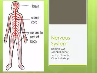
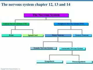
CHAPTER # 13(a). THE PERIPHERAL NERVOUS SYSTEM & REFLEX ACTIVITY. Peripheral Nervous System (PNS). All neural structures outside the brain Sensory receptors Peripheral nerves and associated ganglia Motor endings. Central nervous system (CNS). Peripheral nervous system (PNS).

E N D
CHAPTER # 13(a) THE PERIPHERAL NERVOUS SYSTEM & REFLEX ACTIVITY
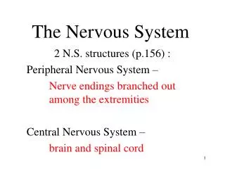
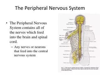
Peripheral Nervous System (PNS) • All neural structures outside the brain • Sensory receptors • Peripheral nerves and associated ganglia • Motor endings
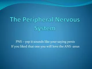
Central nervous system (CNS) Peripheral nervous system (PNS) Sensory (afferent) division Motor (efferent) division Somatic nervous system Autonomic nervous system (ANS) Sympathetic division Parasympathetic division Figure 13.1
Sensory Receptors • Specialized to respond to changes in their environment (stimuli) • Activation results in graded potentials that trigger nerve impulses • Sensation (awareness of stimulus) and perception (interpretation of the meaning of the stimulus) occur in the brain
Classification of Receptors • Based on: • Stimulus type • Location • Structural complexity
Classification by Stimulus Type • Mechanoreceptors—respond to touch, pressure, vibration, stretch, and itch • Thermoreceptors—sensitive to changes in temperature • Photoreceptors—respond to light energy (e.g., retina) • Chemoreceptors—respond to chemicals (e.g., smell, taste, changes in blood chemistry) • Nociceptors—sensitive to pain-causing stimuli (e.g. extreme heat or cold, excessive pressure, inflammatory chemicals)
Classification by Location • Exteroceptors • Respond to stimuli arising outside the body • Receptors in the skin for touch, pressure, pain, and temperature • Most special sense organs
Classification by Location • Interoceptors (visceroceptors) • Respond to stimuli arising in internal viscera and blood vessels • Sensitive to chemical changes, tissue stretch, and temperature changes
Classification by Location • Proprioceptors • Respond to stretch in skeletal muscles, tendons, joints, ligaments, and connective tissue coverings of bones and muscles • Inform the brain of one’s movements
Classification by Structural Complexity • Complex receptors (special sense organs) • Vision, hearing, equilibrium, smell, and taste (Chapter 15) • Simple receptors for general senses: • Tactile sensations (touch, pressure, stretch, vibration), temperature, pain, and muscle sense • Unencapsulated (free) or encapsulated dendritic endings
Unencapsulated Dendritic Endings • Thermoreceptors • Cold receptors (10–40ºC); in superficial dermis • Heat receptors (32–48ºC); in deeper dermis
Unencapsulated Dendritic Endings • Nociceptors • Respond to: • Pinching • Chemicals from damaged tissue • Temperatures outside the range of thermoreceptors • Capsaicin
Unencapsulated Dendritic Endings • Light touch receptors • Tactile (Merkel) discs • Hair follicle receptors
Encapsulated Dendritic Endings • All are mechanoreceptors • Meissner’s (tactile) corpuscles—discriminative touch • Pacinian (lamellated) corpuscles—deep pressure and vibration • Ruffini endings—deep continuous pressure • Muscle spindles—muscle stretch • Golgi tendon organs—stretch in tendons • Joint kinesthetic receptors—stretch in articular capsules
From Sensation to Perception • Survival depends upon sensation and perception • Sensation: the awareness of changes in the internal and external environment • Perception: the conscious interpretation of those stimuli
Sensory Integration • Input comes from exteroceptors, proprioceptors, and interoceptors • Input is relayed toward the head, but is processed along the way
Sensory Integration • Levels of neural integration in sensory systems: • Receptor level—the sensor receptors • Circuit level—ascending pathways • Perceptual level—neuronal circuits in the cerebral cortex
Perceptual level(processing in cortical sensory centers) 3 Motor cortex Somatosensory cortex Thalamus Reticular formation Cerebellum Pons Medulla Circuit level (processing in ascending pathways) 2 Spinal cord Free nerve endings (pain, cold, warmth) Muscle spindle Receptor level (sensory reception and transmission to CNS) 1 Joint kinesthetic receptor Figure 13.2
Processing at the Receptor Level • Receptors have specificity for stimulus energy • Stimulus must be applied in a receptive field • Transduction occurs • Stimulus energy is converted into a graded potential called a receptor potential
Processing at the Receptor Level • In general sense receptors, the receptor potential and generator potential are the same thing stimulus receptor/generator potential in afferent neuron action potential at first node of Ranvier
Processing at the Receptor Level • In special sense organs: stimulus receptor potential in receptor cell release of neurotransmitter generator potential in first-order sensory neuron action potentials (if threshold is reached)
Adaptation of Sensory Receptors • Adaptation is a change in sensitivity in the presence of a constant stimulus • Receptor membranes become less responsive • Receptor potentials decline in frequency or stop
Adaptation of Sensory Receptors • Phasic (fast-adapting) receptors signal the beginning or end of a stimulus • Examples: receptors for pressure, touch, and smell • Tonic receptors adapt slowly or not at all • Examples: nociceptors and most proprioceptors
Processing at the Circuit Level • Pathways of three neurons conduct sensory impulses upward to the appropriate brain regions • First-order neurons • Conduct impulses from the receptor level to the second-order neurons in the CNS • Second-order neurons • Transmit impulses to the thalamus or cerebellum • Third-order neurons • Conduct impulses from the thalamus to the somatosensory cortex (perceptual level)
Processing at the Perceptual Level • Identification of the sensation depends on the specific location of the target neurons in the sensory cortex • Aspects of sensory perception: • Perceptual detection—ability to detect a stimulus (requires summation of impulses) • Magnitude estimation—intensity is coded in the frequency of impulses • Spatial discrimination—identifying the site or pattern of the stimulus (studied by the two-point discrimination test)
Main Aspects of Sensory Perception • Feature abstraction—identification of more complex aspects and several stimulus properties • Quality discrimination—the ability to identify submodalities of a sensation (e.g., sweet or sour tastes) • Pattern recognition—recognition of familiar or significant patterns in stimuli (e.g., the melody in a piece of music)
Perceptual level(processing in cortical sensory centers) 3 Motor cortex Somatosensory cortex Thalamus Reticular formation Cerebellum Pons Medulla Circuit level (processing in ascending pathways) 2 Spinal cord Free nerve endings (pain, cold, warmth) Muscle spindle Receptor level (sensory reception and transmission to CNS) 1 Joint kinesthetic receptor Figure 13.2
Perception of Pain • Warns of actual or impending tissue damage • Stimuli include extreme pressure and temperature, histamine, K+, ATP, acids, and bradykinin • Impulses travel on fibers that release neurotransmitters glutamate and substance P • Some pain impulses are blocked by inhibitory endogenous opioids
Structure of a Nerve • Cordlike organ of the PNS • Bundle of myelinated and unmyelinated peripheral axons enclosed by connective tissue
Structure of a Nerve • Connective tissue coverings include: • Endoneurium—loose connective tissue that encloses axons and their myelin sheaths • Perineurium—coarse connective tissue that bundles fibers into fascicles • Epineurium—tough fibrous sheath around a nerve
Axon Myelin sheath Endoneurium Perineurium Epineurium Fascicle Blood vessels (b) Figure 13.3b
Classification of Nerves • Most nerves are mixtures of afferent and efferent fibers and somatic and autonomic (visceral) fibers • Pure sensory (afferent) or motor (efferent) nerves are rare • Types of fibers in mixed nerves: • Somatic afferent and somatic efferent • Visceral afferent and visceral efferent • Peripheral nerves classified as cranial or spinal nerves
Ganglia • Contain neuron cell bodies associated with nerves • Dorsal root ganglia (sensory, somatic) (Chapter 12) • Autonomic ganglia (motor, visceral) (Chapter 14)
Regeneration of Nerve Fibers • Mature neurons are amitotic • If the soma of a damaged nerve is intact, axon will regenerate • Involves coordinated activity among: • Macrophages—remove debris • Schwann cells—form regeneration tube and secrete growth factors • Axons—regenerate damaged part • CNS oligodendrocytes bear growth-inhibiting proteins that prevent CNS fiber regeneration
Endoneurium Schwann cells 1 The axon becomes fragmented at the injury site. Droplets of myelin Fragmented axon Site of nerve damage Figure 13.4 (1 of 4)
Macrophages clean out the dead axon distal to the injury. 2 Schwann cell Macrophage Figure 13.4 (2 of 4)
Axon sprouts, or filaments, grow through a regeneration tube formed by Schwann cells. 3 Aligning Schwann cells form regeneration tube Fine axon sprouts or filaments Figure 13.4 (3 of 4)
The axon regenerates and a new myelin sheath forms. 4 Site of new myelin sheath formation Schwann cell Single enlarging axon filament Figure 13.4 (4 of 4)