Download
1 / 1
50 likes | 276 Views
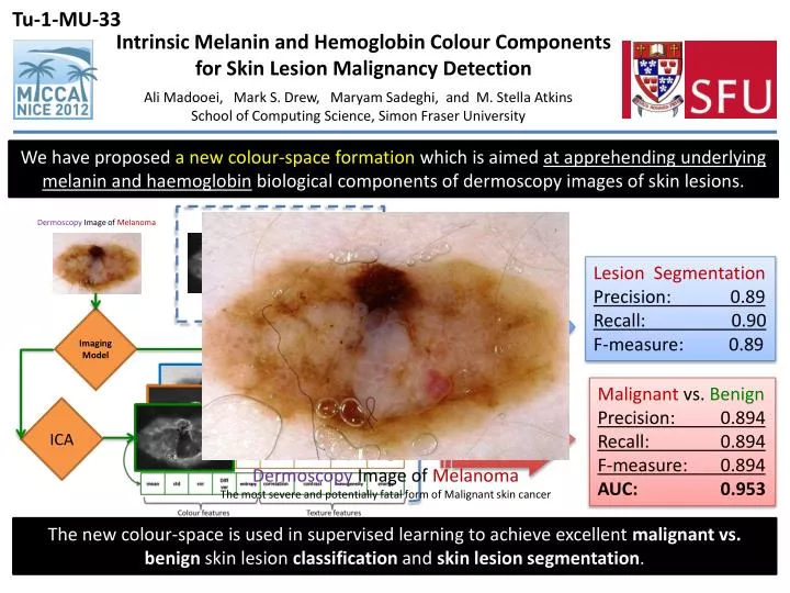
Intrinsic Melanin and Hemoglobin Colour Components for Skin Lesion Malignancy Detection. Ali Madooei, Mark S. Drew, Maryam Sadeghi , and M . Stella Atkins School of Computing Science, Simon Fraser University.

E N D
Intrinsic Melanin and Hemoglobin Colour Components for Skin Lesion Malignancy Detection Ali Madooei, Mark S. Drew, Maryam Sadeghi, and M. Stella Atkins School of Computing Science, Simon Fraser University We have proposed a new colour-space formation which is aimed at apprehending underlying melanin and haemoglobinbiological components of dermoscopy images of skin lesions. We have proposed a new colour-space formation which is aimed at apprehending underlying melanin and hemoglobin biological components of dermoscopy images of skin lesions. Separation of underlying Geo-mean Lesion Segmentation Precision: 0.89 Recall: 0.90 F-measure: 0.89 Dermoscopy Image of Melanoma Imaging Model ICA Otsu Hemoglobin Melanin Malignant vs. Benign Precision: 0.894 Recall: 0.894 F-measure: 0.894 AUC: 0.953 Feature Extraction Logistic Classifier Dermoscopy Image of MelanomaThe most severe and potentially fatal form of Malignant skin cancer The new colour-space is used in supervised learning to achieve excellent malignant vs. benign skin lesion classification and skin lesion segmentation. The new colour-space is used in supervised learning to achieve excellent malignant vs. benign skin lesion classification and skin lesion segmentation.