Download
1 / 31

E N D
1. Cutaneous Malignancy(Nonmelanoma Skin Cancer) UTMB Grand Rounds Presentation:
January 21, 2004
2. 2
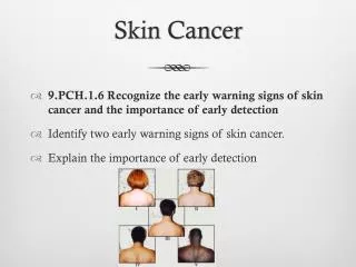
3. 3 Incidence and Epidemiology 800,000 cases per year
Incidence is increasing
Mortality is decreasing
Most occur in patients over 60 years
4. 4 Incidence and Epidemiology - Etiology Ultraviolet light � Sun Exposure
Ionizing radiation causes mutation of tumor suppressor genes
UV B light: 280-320nm, UV A light 320-400nm
Amount of UV B radiation is inversely proportional to ozone
Amount of UV B exposure during childhood and adolescence is directly proportional to risk for BCCA
5. 5 Etiology � Sun Exposure The following groups have the least melanin and are at greatest risk for BCCA:
fair complexion,
light hair,
blue/green eyes,
inability to tan,
history of multiple or severe sunburns,
Celtic ancestry
6. 6 Etiology � Other Factors Arsenic
Radiation Therapy
Burns, Scars, Ulcers
Immunosuppression
Albinism
Bazex's syndrome (basal cell carcinomas, follicular atrophoderma, hypotrichosis, and hypohidrosis or hyperhidrosis)
Gorlin's syndrome (multiple basal cell carcinomas, pitting of the palms and the soles of the feet, mandibular cysts, spine and rib anomalies, calcification of the falx cerebri, and cataracts )
7. 7 Normal Skin Histology
8. 8 Normal Skin Histology Stratum Corneum
Stratum Lucidum
Stratum Granulosum
Stratum Spinosum
Stratum Basale
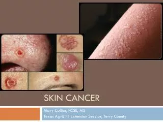
9. 9 Basal Cell Carcinoma Slowly growing malignancy of the epidermis
Rarely metastasizes (.028-.55%)
Cells appear histologically similar to basal cells of epidermis
10. 10 Basal Cell Carcinoma Clinical subtypes
Nodular
Superficial
Pigmented
Morpheaform
11. 11 Basal Cell Carcinoma Nodular
Discrete, raised, circular
Central ulceration
Pink, waxy rolled borders
Relatively non-aggressive
12. 12 Basal Cell Carcinoma Superficial
Threadlike, waxy border
Red, scaling patches
Spread by radial extension
Uncommon in Head and Neck
13. 13 Basal Cell Carcinoma Pigmented
Resemble nevus or melanoma
Behave the same as nodular variant
14. 14 Basal Cell Carcinoma Morpheaform
Macular, whitish, or yellowish plaque
Indistinct clinical margins
15. 15 Basal Cell Carcinoma Histology
Large oval nuclei with little cytoplasm
Nuclei are uniform
Connective tissue stroma causes palisading
16. 16 Basal Cell Carcinoma Histologic Subtypes
Solid
Cystic
Adenoid
Keratotic (Basosquamous)
17. 17 Basal Cell Carcinoma Solid � no cellular differentiation
18. 18 Basal Cell Carcinoma Cystic
Differentiation towards sebaceous glands
Cystic spaces within tumor lobules
19. 19 Basal Cell Carcinoma Adenoid
Glandular pattern
Lacelike pattern
20. 20 Basal Cell Carcinoma Keratotic (Basosquamous)
Basal cell CA with differentiation towards hair structures
Shows feature of both basal cell and squamous cell carcinomas
More aggressive clinically
Undifferentiated cells in combination with parakeratotic cells and horn cysts
21. 21 Squamous Cell Carcinoma More aggressive in terms of local invasion and rate of metastasis than BCCA (2-5%)
Often a progression from sun-damaged areas
Actinic Keratoses
Bowen�s disease
22. 22 Squamous Cell Carcinoma Actinic Keratosis
Indicator of severe sun-damage
<1cm diameter, scaly
Face, scalp, hands, forearms
Progression to SCCA in 20%
Cryotherapy, Shave Excision, 5-FU, TCA
23. 23 Squamous Cell Carcinoma Bowen�s disease
Carcinoma in situ
Well-circumscribed, erythematous scaly patch with irregular border
Common in people with chronic arsenic ingestion
24. 24 Squamous Cell Carcinoma Clinically, SCCA presents as a crusting, erythematous, ulcerated lesion with a granular friable base.
25. 25 Squamous Cell Carcinoma Histology
Irregular masses of epidermal cells proliferating into dermis
Keratinization in well-differentiated tumors
Range in degree of anaplasia
Subtypes of Verrucous, Adenoid squamous, and Spindle Pleomorphic
26. 26 Squamous Cell Carcinoma Histopathology
27. 27 Squamous Cell Carcinoma Verrucous
Minimal atypia
Individual cell keratinization
White, cauliflower lesions
Uncommon in Head and Neck
28. 28 Squamous Cell Carcinoma Spindle-Pleomorphic
Anaplastic
Little keratinization
29. 29 Squamous Cell Carcinoma Adenoid Squamous
Anaplasia
Acantholysis
Tubular and adenoid appearance
30. 30 Squamous Cell Carcinoma
31. 31 Squamous Cell Carcinoma
32. 32 Management Initial evaluation involves
Assessment of location
Punch or excisional biopsy
Staging
33. 33 Management - Staging
34. 34 Management - Curettage Curettes used to remove tumor by feel with small margin of normal tissue
After several cycles, wound is treated topically
Reserved for histologically and clinically favorable basal cell carcinomas.
Not used for squamous cell lesions
35. 35 Management - Cryosurgery Cryogen such as liquid Nitrogen to kill tumor cells
Typical temperature of -50�C .
Tissue-sparing, but leave open wound
Hypopigmentation and scarring may result
Limited to favorable small lesions with well-defined borders
36. 36 Management � Radiation Therapy Used extensively in the past, now sparingly
High cure rate (95%)
Does not allow surgical staging
Protracted treatment course, and expensive
Radiodermatitis, delayed carcinogenesis
Currently reserved for poor operative candidates, adjuvant in high risk malignancy
37. 37 Photodynamic Therapy Photosensitizing drug (porphyrin, 5-ALA) applied topically, orally or parenterally and localizes into tumor cells
Drug is activated by exposure to light (laser)
Efficacy is low (45%)
Side effects include local edema, erythema, blistering, ulceration
Used as palliation
38. 38 Management - Excisional Surgery Most often used by surgeons, esp for larger lesions
Can be with cold steel or laser
Can allow reconstruction in the same sitting
Frozen sections decrease recurrence rate
Can be time consuming and inconvenient
If more than 1/3 of a cosmetic facial unit is excised, better cosmesis with removal of entire unit
39. 39 Management � Excisional Surgery
40. 40 Mohs Surgery - Indications Recurrent skin cancer
Skin cancer in �high risk anatomic areas� and cosmetically important areas
Histologically aggressive skin cancer
Large skin cancers
Skin cancer with ill-defined clinical margins
Irradiated skin
Dermatofibrosarcoma Protuberans
Selected mucosal squamous cell cancers
41. 41 Lymphatic Dissection No hard and fast rules governing lymphatic dissection in N0 Necks
Elective Parotidectomy for deeply invasive tumors of the periauricular region
Large SCCA (>2cm), recurrent SCCA, Marjolin�s ulcer, perineural invasion may require regional lymphadenectomy
Sentinel Lymph Node Dissection may be useful
42. 42 Lymphatic Dissection
43. 43 Merkel�s Cell Carcinoma Tumor of presumed mechanoreceptor origin arising in dermis
Poorly differentiated histology
High rate of recurrence and lymph node metastasis requires excisional surgery with adjuvant radiation and treatment of lymphatic drainage in most cases
44. 44 Merkel�s Cell Carcinoma solitary erythematous to deep purple plaque or nodule of up to several centimeters in size
45. 45 Merkel�s Cell Carcinoma Histology - small, round, basophilic cells arranged in sheets, rests, or trabeculae
Stains for cytokeratins 8, 18, 20
46. 46 Other Rare Cutaneous Malignancies Dermatofibrosarcoma Protuberans
Arises in dermis, spreads deeply
Large indurated plaque with firm irregular flesh colored nodules
Mohs is treatment of choice
Pilomatrix Carcinoma
Arises from Pilomatricoma, a benign tumor of hair matrix origin
Aggressive wide local excision is treatment
47. 47 Conclusions Incidence and Epidemiology
Normal Skin Histology
Basal Cell Carcinoma
Squamous Cell Carcinoma
Treatment of Cutaneous Malignancy
Rare Cutaneous Malignancies
48. 48 Bibliography
25. Lo JS, Snow SN, Reizner GT, Mohs FE, Larson PO, Hruza GJ. Metastatic basal cell carcinoma: report of twelve cases with a review of the literature. J Am Acad Dermatol 1991;24: 715-9.
Sassmannshausen, MD et al �Pilmatrix carcinoma: A report of a case arising from a previously excised pilomatrixoma and a review of the literature,� J Am Acad Dermatol 2001;44:358-61.
Geh JL et al �Unusual multiple pilomatrixomata: case report and review of the literature,� British Journal of Plastic Surgery. 1999; 52(4):320-1
Chih-Shan Jason Chen, �Dermatofibrosarcoma Protuberans,� emedicine.com, October 30, 2003.
Swanson, NA, �Mohs surgery: technique, indications, applications, and the future.� Arch Dermatol 1983; 1, 19:761.
Boone, John L, �Merkel Cell Tumors of the Head and Neck,� emedicine.com, September 8, 2003
Stucker, Fred J. �Cutaneous Malignancy,� Bailey, Byron J. Head & Neck Surgery � Otolaryngology, Lippincott Williams and Wilkins, Philadelphia, 2001.