Download
1 / 1
10 likes | 143 Views
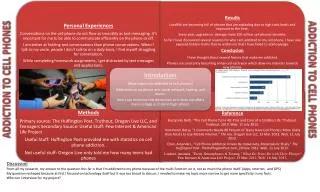
siRNA-D5. A. 3.9. B. 1.6. ns-siRNA. 3.6. 1.3. untreated. 3.5. 0.8. 24 – 72 h. 20 h. 24 – 72 h. WST-1 assay cell counting apoptosis detection cell cycle distribution. CT (24 h). seeding. transfection (4 h). 24 – 72 h. 20 – 68 h. WST-1 assay cell counting RNA isolation

E N D
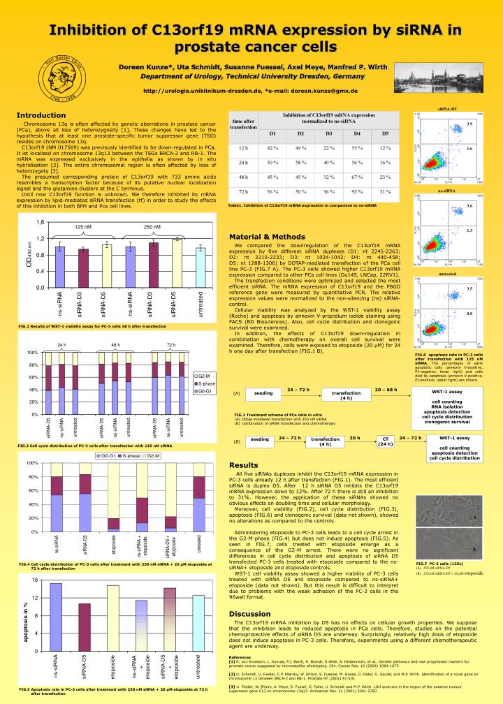
siRNA-D5 A 3.9 B 1.6 ns-siRNA 3.6 1.3 untreated 3.5 0.8 24 – 72 h 20 h 24 – 72 h WST-1 assay cell counting apoptosis detection cell cycle distribution CT (24 h) seeding transfection (4 h) 24 – 72 h 20 – 68 h WST-1 assay cell counting RNA isolation apoptosis detection cell cycle distribution clonogenic survival seeding transfection (4 h) (A) FIG.7 PC-3 cells (125x) (A) 250 nM siRNA-D5 (B) 250 nM siRNA-D5 + 20 µM etoposide (B) FIG.6 apoptosis rate in PC-3 cells after transfection with 125 nM siRNA. The percentages of early apoptotic cells (annexin V-positive, PI-negative; lower right) and cells died by apoptosis (annexin V-positive, PI-positive; upper right) are shown. • Inhibition of C13orf19 mRNA expression by siRNA in prostate cancer cells • Doreen Kunze*, Uta Schmidt, Susanne Fuessel, Axel Meye, Manfred P. Wirth • Department of Urology, Technical University Dresden, Germany http://urologie.uniklinikum-dresden.de, *e-mail: doreen.kunze@gmx.de Introduction Chromosome 13q is often affected by genetic aberrations in prostate cancer (PCa), above all loss of heterozygosity [1]. These changes have led to the hypothesis that at least one prostate-specific tumor suppressor gene (TSG) resides on chromosome 13q. C13orf19 (NM 017569) was previously identified to be down-regulated in PCa. It ist localized on chromosome 13q13 between the TSGs BRCA-2 and RB-1. The mRNA was expressed exclusively in the epithelia as shown by in situ hybridization [2]. The entire chromosomal region is often affected by loss of heterozygoty [3]. The presumed corresponding protein of C13orf19 with 733 amino acids resembles a transcription factor because of its putative nuclear localization signal and the glutamine clusters at the C terminus. Until now C13orf19 function is unknown. We therefore inhibited its mRNA expression by lipid-mediated siRNA transfection (tf) in order to study the effects of this inhibition in both BPH and Pca cell lines. Table1. Inhibition of C13orf19 mRNA expression in comparison to ns-siRNA Material & Methods We compared the downregulation of the C13orf19 mRNA expression by five different siRNA duplexes (D1: nt 2245‑2263; D2: nt 2215‑2233; D3: nt 1024‑1042; D4: nt 440‑458; D5: nt 1288‑1306) by DOTAP-mediated transfection of the PCa cell line PC-3 (FIG.7 A). The PC-3 cells showed higher C13orf19 mRNA expression compared to other PCa cell lines (Du145, LNCap, 22RV1). The transfection conditions were optimized and selected the most efficient siRNA. The mRNA expression of C13orf19 and the PBGD reference gene were measured by quantitative PCR. The relative expression values were normalized to the non-silencing (ns) siRNA-control. Cellular viability was analyzed by the WST‑1 viability assay (Roche) and apoptosis by annexin V-propidium iodide staining using FACS (BD Biosciences). Also, cell cycle distribution and clonogenic survival were examined. In addition, the effects of C13orf19 down-regulation in combination with chemotherapy on overall cell survival were examined. Therefore, cells were exposed to etoposide (20 µM) for 24 h one day after transfection (FIG.1 B). FIG.2 Results of WST‑1 viability assay for PC-3 cells 48 h after transfection • FIG.1 Treatment scheme of PCa cells in vitro • Dotap-mediated transfection with 250 nM siRNA • combination of siRNA transfection and chemotherapy FIG.3 Cell cycle distribution of PC-3 cells after transfection with 125 nM siRNA Results All five siRNAs duplexes inhibit the C13orf19 mRNA expression in PC-3 cells already 12 h after transfection (FIG.1). The most efficient siRNA is duplex D5. After 12 h siRNA D5 inhibits the C13orf19 mRNA expression down to 12%. After 72 h there is still an inhibition to 31%. However, the application of these siRNAs showed no obvious effects on doubling time and cellular morphology. Moreover, cell viability (FIG.2), cell cycle distribution (FIG.3), apoptosis (FIG.6) and clonogenic survival (data not shown), showed no alterations as compared to the controls. Administering etoposide to PC-3 cells leads to a cell cycle arrest in the G2-M-phase (FIG.4) but does not induce apoptosis (FIG.5). As seen in FIG.7, cells treated with etoposide enlarge as a consequence of the G2-M arrest. There were no significant differences in cell cycle distribution and apoptosis of siRNA D5 transfected PC-3 cells treated with etoposidecompared to the ns-siRNA+ etoposide and etoposidecontrols. WST-1 cell viability assay showed a higher viability of PC-3 cells treated with siRNA D5 and etoposidecompared to ns-siRNA+ etoposide (data not shown). But this result is difficult to interpret due to problems with the weak adhesion of the PC-3 cells in the 96well format. FIG.6 clonogenic survival, PC-3, 125 nM siRNA FIG.4 Cell cycle distribution of PC-3 cells after treatment with 250 nM siRNA + 20 µM etoposide at 72 h after transfection Discussion The C13orf19 mRNA inhibition by D5 has no effects on cellular growth properties. We suppose that the inhibition leads to reduced apoptosis in PCa cells. Therefore, studies on the potential chemoprotective effects of siRNA D5 are underway. Surprisingly, relatively high dosis of etoposide does not induce apoptosis in PC-3 cells. Therefore, experiments using a different chemotherapeutic agent are underway. References [1] R. von Knobloch, L. Konrad, P.J. Barth, H. Brandt, S.Wille, A. Heidenreich, et al., Genetic pathways and new progression markers for prostate cancer suggested by microsatellite allelotyping. Clin. Cancer Res. 10 (2004) 1064-1073 [2] U. Schmidt, U. Fiedler, C.P. Pilarsky, W. Ehlers, S. Fuessel, M. Haase, G. Faller, G. Sauter, and M.P. Wirth. Identification of a novel gene on chromosome 13 between BRCA-2 and RB-1. Prostate 47 (2001) 91-101 [3] U. Fiedler, W. Ehlers, A. Meye, S. Fussel, G. Faller, U. Schmidt and M.P. Wirth. LOH analyses in the region of the putative tumour suppressor gene C13 on chromosome 13q13. Anticancer Res. 21 (2001) 2341-2350 FIG.5 Apoptosis rate in PC-3 cells after treatment with 250 nM siRNA + 20 µM etoposide at 72 h after transfection