Download
1 / 1
10 likes | 141 Views
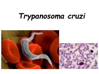
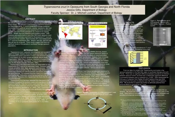
30. 25. 20. 2003. 15. 2004. 10. 5. 0. Trypanosoma cruzi in Opossums from South Georgia and North Florida Jessica Gillis, Department of Biology Faculty Sponsor: Dr. J. Mitchell Lockhart, Department of Biology. ABSTRACT

E N D
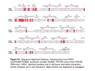
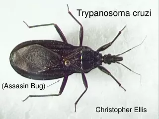
30 25 20 2003 15 2004 10 5 0 Trypanosomacruzi in Opossums from South Georgia and North Florida Jessica Gillis, Department of Biology Faculty Sponsor: Dr. J. Mitchell Lockhart, Department of Biology ABSTRACT Trypanosomacruzi is a protozoan parasite causing a disease that affects many in Central and South America, but only few in the United States. The disease is transmitted by triatomine insects of the Reduviidae family. T.cruzi is predicted to occupy numerous mammalian hosts such as raccoons, coyotes, opossums, deer and dogs. Over 1,000 opossums were acquired form three southwest Georgia and north Florida plantations as a result of an ongoing USDA – Wildlife Services bobwhite-quail predator project. DNA isolations were performed on more than 200 frozen opossum heart tissue samples and were examined for the presence of T. cruzi via polymerase chain reaction. Approximately 20% have tested positive for T. cruzi as of submission date. Season, host sex, and host age data will also be analyzed. These results suggest that there are significant levels of this parasite in southwest Georgia opossum populations. Figure 4. Representative gel of PCR products. Bottom Lane – DNA ladder, Top Lane – Positive Control RESULTS From 2003 and 2004, two-hundred and fourteen opossums samples were tested for Trypanosoma cruzi and 42 (19.6%) were positive (see Figure 4 for representative gel). From 2003, 15/122 opossums (12.3%) from Pebble Hill and Pinebloom-East Plantations were positive for T. cruzi. 27/92 (29.3%) opossums from Tall Timbers and Pinebloom-West plantations tested positive for T. cruzi in 2004. 29/127 (22.8%) of female opossums and 13/87 (14.9%) of male opossums were positive. Figure 1. Worldwide distribution of Chagas Disease Figure 2. Trypanosomacruzi life cycle Figure 5. Yearly prevalence of T. cruzi in opossums. Prevalence is on y axis. METHODS The United States Department of Agriculture-Wildlife Services, Auburn University, The University of Georgia, and Tall Timbers Research Station in Florida began a project in 2001 to monitor the effects of mesomammalian predator removal on bobwhite quail reproduction. Valdosta State University became a research collaborator on the project in 2003. Predators were removed from Pebble Hill Plantation, Pinebloom Plantation and Tall Timbers Research Station by the USDA-WS. Animals were euthanized, frozen and transported to VSU for study. Predators included opossums, raccoons, foxes, armadillos, coyotes, feral dogs, feral cats, and bobcats. Animals were necropsied and tissue samples were collected. Various natural history parameters were recorded. Two-hundred and fourteen frozen heart tissue samples were examined for the presence of T. cruzi using polymerase chain reaction. The frozen samples were thawed and DNA isolations were performed using Qiagen DNeasy™ tissue kits following the manufacturer’s protocol. PCR, using T .cruzi specific primers MCS-35 and MCS-36 found in a 330 base pair region of the kinetoplast DNA minicircle of T .cruzi, was performed (Figure 3). Products of PCR were electrophoresed in 2% agarose gels and visualized using ethidium bromide and photographed for analysis (Figure 4). Positive and negative controls were run for each set of samples. Precautions were taken and strict protocols were followed to prevent contamination. INTRODUCTION Trypanosoma cruzi, a parasitic hemoflagellate and causative agent of Chagas’ disease (American trypanosomiasis), has devastated the human population in many Latin American countries. T. cruzi infects 16-18 million people worldwide (World Health Organization, 1993) (Fig.1), however, infections in humans in the United States are rare. Worldwide, T. cruzi is responsible from more than 50,000 deaths annually (Tanowitz, 1992). Kissing bugs in the Reduviidae family are responsible for the transmission of T. cruzi to its mammalian hosts. Common hosts include armadillos, deer, dogs, opossums, and raccoons. The life cycle of T. cruzi is complex (Fig. 2). Assassin bugs feed on the blood of an infected host and the parasite reproduces asexually in the gut of the bug. The assassin bug then feeds on a new host by piercing the skin. Defecation occurs during feeding on the skin of the new host. Parasites then migrate from the feces into the open wound of the host. Trypanosoma cruzi has been detected from Maryland in the northeast throughout the southern states and extending to the west coast of California. Only five human cases have been reported in the United States and have come from California, Tennessee, and Texas (reviewed by Herwakdt et. al., 2000). The T. cruzi strain that exists in the United States seems to be significantly less pathogenic than that found in South America. The Virginia opossum (Didelphisvirginianus) is a frequent carrier of Trypanosoma cruzi. In previous studies, 89 of 552 (16%) opossums tested in Georgia and northwest Florida were positive for T. cruzi (McKeever et al., 1958). In North Carolina, 1 of 12 (8.3%) opossums were carriers of T. cruzi (Karsten et al., 1992). In Louisiana, 37.5% of opossums were positive for T. cruzi (Barr, 1991). DISCUSSION From our data it is evident that Trypanosoma cruzi exists in the opossum population in the Red Hills region of southwest Georgia and north Florida. There was no significant difference between prevalence of infection between opossum sexes, but there was a statistically significant difference in prevalence between year of collection (p > 0.005). 2003 represented the third year of collection from Pebble Hill and Pinebloom-East and 2004 represented the first year of collection from Tall Timbers and Pinebloom-West. We would like to explore this further to delineate potential host removal effects on prevalence of T. cruzi. FUTURE RESEARCH In the future, we would like to incorporate specific age data into our analysis. We would also like to evaluate data from the final two years of the study. Other factors to consider include habitat management strategies and possibly to incorporate existing geographic information systems data. Figure 3. Schematic representation of a kDNA minicircle of T. cruzi Arrows indicate the relative hybridization locations of the MCS35 and MCS36 primers, which give an amplification product of 330bp MCS36 REFERENCES Barr, S.C., C.C. Brown, V.A. Dennis, and T.R. Klei. 1991. The Lesions and Prevalence of Trypanosoma cruzi in Opossums and Armadillos from Southern Louisiana. Journal of Parasitology 77: 624- 627. Herwaldt, B. L., M. J. Grijalva, A. L. Newsome, C.R. McGhee, M. R. Powell, D. G. Nemec, F. J. Steurer, and M. L. Eberhard. 2000. Use of polymerase chain reaction to diagnose the fifth reported U. S. case of autochthonous transmission of Trypanosoma cruzi in Tennessee, 1998. Journal of Infectious Diseases 181: 395-399. Karsten, V., C. Davis, and R. Kuhn. 1992. Trypanosoma crusi in wild raccoons and opossums in North Carolina. Journal of Parasitology 78: 547-549. McKeever, S., G.W. Gorman, and L. Norman. 1958. Occurrence of Trypanosoma cruzi-like organism in some mammals from southwestern Georgia and northwestern Florida. Journal of Parasitology 44: 583-587. Tanowitz, H. B., L. V. Kirchhoff, D. Simon, S. A. Morris, L. Weiss, and M. Wittner. 1992. Chagas disease. Clinical Microbiology Review S: 400-419. World Health Organization. 1993. Chagas’ disease. In Tropical diseases. Tropical disease research. Progress 1991-1992. Eleventh Programme Report of the UNDP/World Bank/WHO Special Program for Research and Training in Tropical Diseases, Geneva, Switzerland, p. 67-75. MCS35 MCS36 MCS35 MCS36 MCS35 MCS35 MCS36 330 bp