Download
1 / 44
590 likes | 1.04k Views
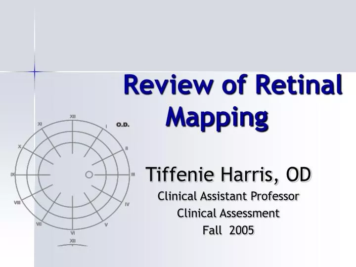
Review of Retinal Mapping. Tiffenie Harris, OD Clinical Assistant Professor Clinical Assessment Fall 2005. Review of BIO Technique. Sitting vs. Supine Sitting allows folds within the RD or break to open Smooth and efficient

E N D
Review of Retinal Mapping Tiffenie Harris, OD Clinical Assistant Professor Clinical Assessment Fall 2005
Review of BIO Technique • Sitting vs. Supine • Sitting allows folds within the RD or break to open • Smooth and efficient • Systematically examine the quadrants out to the periphery • “Sweeping” views of retina • Posterior pole last • Patient head position • Stand/sit directly opposite meridian of interest • Turn the patient’s head toward the examiner/ have the patient look toward the meridian of interest • Illumination • Light fundus vs. dark • Periphery vs. central
Review of Anatomical Landmarks • Horizontal meridian • Long posterior ciliary nerves at 3 & 9 o’clock represent the anatomical horizontal meridians • divides the retina into superior/inferior • Vertical meridian • Short ciliary nerves seen between 10 and 2 as well as 4 and 6 o’clock represent the anatomical vertical meridians • Divides the retina into nasal/temporal
Long Posterior Ciliary Nerve • Retinal photo of patient looking left with OD
Review of anatomical landmarks • Anterior/Posterior retina is divided by the Equator • Vortex ampulla are located just posterior(approx 3mm)and there are usually 4 to 6 vortex veins • located in the 1, 5, 7, 11 o’clock meridians • Ora Serrata • The junction between the retina and ciliary body • Nasal ora appears “serrated” and the temperal does not • Posterior pole • Macula and optic nerve along with the sup/inf vessel arcades
Ora Serrata • Nasal or Temperal retina?
Extended OphthalmoscopyCPT 92225 • Documentation requirements • A detailed sketch must be included in the medical record with sufficient labeling along with an interpretation that affects the plan of treatment • The sketch should be a minimum size of 3-4" in diameter • All items noted must be identified • Drawings in 4-6 standard colors are preferred • 92225-Extended ophthalmoscopy is one of the most heaviest audited codes by medicare
Limitations • Considered not medically necessary if • there is insufficient drawing • the medical record does not document the interpretation • it replaces a routine ophthalmoscopy. • when other testing such as fundus photography gives the same information • It is not enough to link the procedure code to a correct, payable ICD-9-CM code • The diagnosis or clinical signs/symptoms must be present for the procedure to be paid.
Conventional Color Scheme for Retinal Mapping -Red: Light red: attached retina Dark red: retinal arteries, preretinal or intraretinal hemorrhages -Light blue: Retinal Detachment -Dark blue: Retinal veins, margins of retinal breaks, lattice is outlined in blue with inside crosslined --Black: chorioretinal pigmentation -Yellow: intraretinal or subretinal exudates -Brown: nevi, melanomas, choriodal detachment -Green: vitreous or lens opacities Opacities in media, vitreous hemorrhage
Example #1 28 yo 8D myopic female Cc: floaters/flashes x 3-4 days, no hx of injury DVA: 20/20 OD, 20/20 OS PERRLA, EOM: snf, Slex: C/C- clr, L/L –clr AC: C&Q, VH-Gd4
Example #1 • What additional tests/examination can you perform? • What do you expect to find? • Why are her acuities good? • Would you expect her to have an APD?
Example #1 • CVF: FTFC – OS, superior defect noted OD, -APD • +Shaffer’s sign • The presence of pigment granules in the anterior vitreous (Berger's space) AKA "tobacco dust," • Very significant clues to a retinal break or detachment. Hamilton and Taylor found that 98% of patients in their clinical review with this sign had retinal detachments and 60% had flat retinal holes • Failure to check for this sign on a symptomatic patient could be considered gross negligence • Red blood cells, secondary to a vitreous hemorrhage, may be difficult to differentiate from the pigment granules. However, with a red free filter (green) the red blood cells will disappear The pigment granules will not absorb the red-free light and will still be seen.
Example #1-OD looking down and right -What do you see? -Where do you draw your findings?
ok • VR consultants
Where do you draw your findings? Example #2-OS looking up and left
Where do you draw your findings? Example #3-OD looking up and right
Sketch What You See Assignment Due: October 5th Clinical Assessment Fall 2005
Instructions • Use stencils if available • Record locations by clock hour when appropriate • Include anatomical landmarks to reference location • Use labels with arrows when appropriate • Approximate size of lesion (range is allowed) use optic disc as gauge (e.g., 2DD in size) • Note topographical characteristics when appropriate (e.g., elevations, escavations, etc.) • Each item is worth 10 points of credit and partial credit will be given, 100 total points possible
This is a 20D view of the posterior pole Which eye is this? Provide a list of differentials for this retinal finding (at least 5) Case #1
Case #2 This small circle is an artifact
This is a retinal photo of a patient looking up and to the left with the right eye What is this retinal finding? Provide a differential list for this retinal finding (list at least 3 conditions) Case #2
This is a 20D view of OS looking up and to the right What is this retinal finding? What symptoms do you expect this patient to experience? Case #3
Describe what you see in this retinal photo as you would record it in a patient’s record Provide a list of differentials for this fundus finding What is your best answer and why? Case #4
Case #5 • This is a retinal photo of OD looking up • Provide a list of differentials for this retinal finding (at least 5) Bonus: What is this retinal finding?
This is a 20D view of OD looking down to the left What is this retinal finding? What are the risks associated with this finding? Case #6
Case #7 • The white spots are artifacts on the photo
Describe what you see in this retinal photo as you would document it in a patient’s record What is the retinal finding? Provide a list of differentials (at least 3) BONUS: What tests should be performed on this patient? Case #7
This is a 20D view of OD looking down Provide a differential list for this retinal finding (at least 3) What is your answer and why? Case #8
This is a retinal photo of OD looking right and slightly down Provide a list of differentials for this retinal finding(at least 3) What symptoms might this patient experience? Case #9
Case #10 • What is this fundus finding seen in this retinal photo? • This diabetic patient has what stage of diabetic retinopathy? NOTE: be as specific as possible • Give at least ONE differential
What is this retinal condition? Why does the retina appear this way? Be as specific as possible What is the expected visual acuity? Is there any underlying systemic conditions associated with this retinal finding? Extra Credit Case