Download
1 / 33
340 likes | 572 Views
Retinal Projections. • The type of bipolar cell innervated by 1 cone is a midget bipolar cell. These may be of the “on” or “off” subtype. . A cone transmits glutamate to each, but with incident light it hyperpolarizes and stops transmitting glutamate.

E N D
• The type of bipolar cell innervated by 1 cone is a midget bipolar cell. • These may be of the “on” or “off” subtype.
A cone transmits glutamate to each, but with incident light it hyperpolarizes and stops transmitting glutamate. • “Off” bipolar cells have normal excitatory AMPA receptors, so that with light, no glutamate comes and they are off.
“On” bipolar cells have hyperpolarizing glutamate receptors called mGluR6. • With light, the lack of glutamate leads to depolarization/signaling.
• The surround region is opposite to the center region and is mediated by lateral inhibition from horizontal cells. • Horizontal cells release GABA onto the synapse of the center photoreceptor and its bipolar cell.
• On and Off bipolar cells project to On and Off ganglion cells with the same types of properties. • These ganglion cells project to the brain, though the retina itself is a CNS outgrowth.
• These two types of cells create a situation where any stimulus depolarizes one population of bipolar/ganglion cells and hyperpolarizes another. • • On cells respond to light shining on their centers, or a darker stimulus going away. • Likewise, Off cells respond to darkness on their centers, or a light stimulus being taken away.
• At a border of light/dark, an On ganglion cell on the light side of the border has it’s center in the light and surround in the dark. • So, the darker the dark area, the more that ganglion cells is stimulated and the lighter you interpret the light area.
This produces optical illusions or light and dark. • • Rods mediate vision with light in the scotopic range (very dim), whereas in the mesopic vision you use both, and in the photopic range cones mostly mediate vision.
There are three types of cone pigments and one rod type. Of cone pigments, 60% are red, 30% green, and only 5% blue. • • Each cone only expresses one opsin. • The range of sensitivity of the opsins overlap extensively.
With one type of cone, you couldn’t tell colors apart because you’d never know if the difference in stimulation was due to light intensity of the color of the light. • Multiple cones gives a ratio of activity of the two that is independent of intensity.
• Opsins are the proteins that bind 11-cis-retinal and tune its absorption spectrum. • By changing AAs in the opsin, you can alter the wavelength sensitivity. • The divergence of red and green opsins occurred recently in evolution.
They are adjacent on the X-chromosome, as the result of a gene duplication, and are commonly subject to mitotic errors that produce RG colorblindness.
• The fovea is the point of highest visual acuity. • It represents only 1% of the visual field, but projects to about half of the visual cortex. • It has only cones, which are very small (midget cones). • The other specialization is the optic disc, which has no retinal neurons so produces a blind spot.
80% of the ganglion cells, and contribute to the ability to see shape (due to small RF and center/surround patterning) as well as the ability to differentiate colors.
P-type ganglion cells get input from red and green cones. • The “center” is generated by either 1 red cone or 1 green cone (it may still be On or Off though), and the “surround” is driven by many red and green cones.
• So, not only are the center and surround different in their on and off capabilities, but also they respond best at different wavelengths.
Thus, these cells will respond optimally with only the center illuminated with one color, but will still respond above baseline with their entire RF covered in a wavelength to which the center is more sensitive than the surround.
• There are P and M type ganglion cells. • P are small, with both chromatic and spatial (on/off) contrast. • M ganglion cells are large, have spatial contrast (on/off), but don’t have chromatic contrast. • They have red and green cones in both the center and surround.
In M-cell terminology, they call red “long wavelength” and green “medium wavelength” sensitive. • • M cells are also responsive to movement, and they are rapidly adapting. • They have large RFs, so respond more to depth and movement rather than form.
• Blue cones project to small bistratified ganglion cells. • They have no center/surround organization, rather they tend to be Blue-On and Red/Green-Off. • They have huge RFs and don’t care at all about shape, only about color. • 3 blue cones go to 1 bipolar cell that inputs onto the Blue ganglion cell.
Many red and green cones go to a second bipolar cell that inputs onto the ganglion cell. • • Melanopsin ganglion cells are light sensitive with very large RFs, but don’t contribute to the imaging pathways.
They innervated the suprachiasmatic nucleus (circadian rhythms) and the pretectal nucleus (pupillary constriction). • They spike with the onset and turning-off of light.
• At low light levels there is rod activity, but no cone activity. • But, there’s no dedicated pathway for rods, so they share the same ganglion cells that transmit cone signals.
All rod bipolar cells are “on” with light, and interestingly their bipolar cells don’t directly synapse with ganglion cells: Light → Rod → “On” Bipolar Cell → Amacrine cell AP → electrical synapse to increase the response of an ‘On” ganglion cell, or chemical GABA synapse to decrease the response of an “Off” ganglion cell.
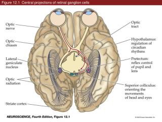
• The nasal half of the retina gives off the axons of the optic nerve that will cross to the contralateral side at the optic chiasm. • This allows for information from one side of the visual field (in both eyes) to go to the same physical location in the brain. • So, each optic tract (after the chiasm) has nasal retinal axons from the contralateral side and temporal retinal axons from the ipsilateral side.
• In animals with laterally placed eyes, all axons cross the midline. • In us, 120 degrees of the 160 degrees of visual field are covered by both eyes. • The crossing pattern in us allows information from one side of the visual field to go to the contralateralhemisphere. • This is also important for steropsis (depth perception).
• The image forming pathway. 90% of axons in the optic nerves go to the LGN of the thalamus, which sends axons to V1 in the occipital lobe. • These include axons of M, P, and small bistratified ganglion cells. • The other 10% are for pupillary constriction (melanopsin ganglion cells go to the pretectum) or visual reflexes (to the superior colliculus).
• The LGN has 6 numbered layers, with 1 most ventral. • Layers 1 and 2 and magnocellular layers with large cells, and layers 3-6 are parvocellular layers with smaller cells. • The LGN keeps information from the contralateral and ipsilateral eyes separate.
Each eye projects to one of the two magnocellular layers and two of the four parvocellular layers of each LGN. • The two parvocellular layers from each eye are separated based on their origin in “on” vs. “off” bipolar cells.
• RFs of LGN cells are very similar to the RFs of the ganglion cells that innervate them, so very little processing/integration occurs here. • They slightly refine RFs. • Their firing is also dependent on your state of arousal and attention.
• The M and P layers of the LGN are driven by red (long λ) and green (med λ) cones. • The regions between layers of the LGN are called Koniocellular layers, which are small and get input from the blue responsive ganglion cells.
• So overall, each ganglion cell population has its own target in the LGN. • • From the LGN, optic radiations carry axons to the primary visual area in the occipital lobe, where images are analyzed.