Download

1 / 62
620 likes | 833 Views
Skin and Soft Tissue Tumors. Dr. Jamaleldin Hassainan. Arise from any histological structures that make up skin. Epidermis Connective tissue Glands Muscle Nerves. CLASSIFICATION. Benign Premalignant Malignant. Common Benign Tumors. Heamangiomas : Involuting Non- involuting.

E N D
Skin and Soft Tissue Tumors Dr. Jamaleldin Hassainan
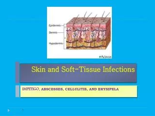
Arise from any histological structures that make up skin • Epidermis • Connective tissue • Glands • Muscle • Nerves
CLASSIFICATION • Benign • Premalignant • Malignant
Common Benign Tumors • Heamangiomas : • Involuting • Non- involuting
Involuting Heamangiomas • Heamangiomas of childhood • 95% of all heamangiomas • Not a true neoplasm • Neoplasm of endothelial cells • Undergo complete spontaneous involution
Involuting Heamangiomas (cont.) • Present at birth or appears 2-3 weeks after birth • Grows rapidly 4-6 months • Spontaneous involution complete 5-7 yrs
Classification Involuting • Superficial • Combined • Deep
Superficial Involuting • Strawberry nevus • Nevus vasculosa • Capillary heamangioma Appearance : • Sharp demarcated red • Slightly raised lesion & irregular surface
COMBINED • Strawberry • Capillary & Cavernous Appearance : A firm bluish tumor , may extend deeply into sub cutaneous surface
Deep Involuting • Cavernous • Appearance : Blue tumor covered by normal skin • Treatment : Requires no treatment involving vital organ eg. lid
Non Involuting Heamangiomas • Usually present at birth • No rapid growth • Growth is proportion to growth of child • Persists into adulthood • Causes severe aesthetic problems • May cause arterio venous fistula , eventually lead to cardiac failure. • Treatment : Not satisfactory
Port Wine Stain • May involve any portion of the body • When present in face as a flat patch correlating to sensory branch of 5th nerve • Microscopic appearance : • Thin walled capillaries distributed throughout the dermis lined by thin mature endothelial cells • Treatment :Unsatisfactory - Tattooing - Laser -Radiotherapy
Malignant Tumors • Basal cell carcinoma • Squamous cell carcinoma • Malignant Melanoma
Basal Cell Carcinoma (Rodent ulcer) • Most common malignant carcinoma • Predisposing factors : • Age >40 yrs • Ultraviolet light exposure • Fair skin , blond hair & blue eyes living in tropical climate i.e. westerners living in Saudi Arabia .
Predisposing Factors (cont.) • Growth is slow , steady & insidious. Several years may pass before patient becomes concerned. • Invade adjacent tissue , massive ulcerations . • Rarely metastases & death may occur by invading deeper extension into intracranial or major blood vessels.
APPEARANCE • Small , translucent skin elevated nodule • Rolled pearly edges • Telangiactic vessels occur commonly on surface
Sclerosing Morphia • Less common • Elongated strands of basal that infiltrate the dermis . • Flat & whitish or waxy appearance and firm palpation
Erythromateous Basal Cell Carcinoma • Body basal occurs most frequently on the trunks. • Appears reddish plaques with atrophic center • Smooth slightly raised borders.
Pigment Basal • Sometimes mistaken for melanoma
Treatment • Radio therapy : Good in treatment of structures that are difficult to reconstruct . Should not be used in pt. under 40 y , or in pt. who failed to respond to radiation therapy Treatment : 4-6 weeks
Treatment • Curettage & Electro desiccation : Excise 2-3 mm margin • Surgical excision : smallmoderate size lesion down to subcutaneous tissue
Squamous Cell Carcinoma • 1st most cancer in dark skinned people • 2nd most cancer in light skinned group • Causative agents same as basal cell carcinoma . • Most common sites are the ears , cheeks , lower lip & back of the hands.
Squamous cell (cont.) • Other causative agents are chronic contact with tars hydrocarbons & exposure to ionizing radiation . • Also chronic ulcers , thermal burns healed with fibrosis ( Marjolins ulcer ) • These are aggressive tumors , does not usually metastasize , as fibrosis & initial burns has already destroyed lymphatic
Presentation • Locally invasive without metastasizing from premalignant tumors eg. Bowens disease , chronic radiation dermatitis. • Rapidly growing widely invasive with metastasizes especially squamous cell tumors arising from normal skin .
Presentation (cont.) • Grows initially starts as a erythomatous plaque or nodule with indistinct margins. • Surface may be : - Flat - Verocous - Ulcerative • Histopathology : Malignant epithelium cell are seen extending down into the dermis like horn pearls . • Treatment : - Surgery-Radiation
Types of Nevi • Junctional Nevi: • Are small , circumscribed , light brown or black , flat – slightly raised & rarely contained hair • Mainly lies between dermis & epidermis these may be found in mucous membrane ,genitalia , soles & palms