Download
1 / 40
850 likes | 5.16k Views
Ankle Arthrodesis. Dr. C Wadden Dr. K-A Lalonde May 3, 2012. Overview. History Anatomy Indications Surgical Options Complications Outcomes. History. Originally described in 1879 by Albert Treatment for TB of ankle joint Over thirty procedures described

E N D
Ankle Arthrodesis • Dr. C Wadden • Dr. K-A Lalonde • May 3, 2012
Overview • History • Anatomy • Indications • Surgical Options • Complications • Outcomes
History • Originally described in 1879 by Albert • Treatment for TB of ankle joint • Over thirty procedures described • Charnley developed compression technique in 1951, used an external-fixator • Arthroscopic arthrodesis described in 1983 • Mini-open arthrodesis described in 1996
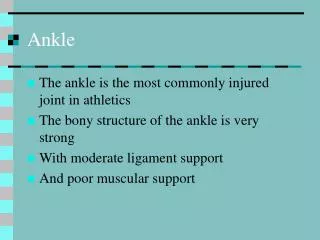
Anatomy • Tibiotalar or talocrural joint is a hinge joint • consists of talar dome, tibial plafond, malleoli • Isolated movement occurs in the sagittal plane • 18o dorsiflexion to 48o plantar flexion • Small amounts of movement in coronal and axial planes • IR/ER in axial plane • Inversion/Eversion in coronal plane
Indications • Principle indication is pain and stiffness that is functionally disabling • prior #, infection, osteonecrosis, osteochondral defects, OA, RA • Absence of arthritis and normal alignment in the subtalar complex • talocalcaneal, talonavicular, calcaneocuboid
Evaluation • History and Physical Examination • Weight bearing AP and Lateral radiographs • CT +/- arthrography • Selective joint injections
Surgical Options • External Fixation • severe osteopenia, pre-existing septic joint • Open arthrodesis • significant deformity or malalignment • Mini-open/Arthroscopic arthrodesis • minimal deformity
Position of Fusion • Optimal ankle position is the same regardless of surgical technique • Ankle • Neutral flexion (0o) • 5o - 10o ER (comparable to contralateral side) • Slight valgus (5o) • Translate talus posteriorly to align with posterior margin of tibia
Position of Fusion • Ankle’s fused in neutral have 10o of plantar flexion though midfoot at heel strike • approximates normal ankle producing relatively normal barefoot gait pattern • Shifting the talus posteriorly + ER of 5o - 10o reduces lever arm of the foot • mild push-off by pronation through subtalar complex • Neutral position best utilizes midfoot motion to simulate normal ankle
External Fixation • Charnley Method • Open debridement of ankle joint cartilage + Ex-fix • External-fixator • One pin through tibia • One pin through neck of talus • Connecting bars • Compression relies on intact achilles tendon
External Fixation • Calandruccio external fixator • triangular, resists torsion, does not require intact achilles • Open debridement, external-fixator placed • Pins through neck and body of talus • Pin through tibia • Occasionally one pin through calcaneus • Fusion site buttressed with bimalleolar onlay grafts
External Fixation • Unilateral external fixator • adequate resistance to dorsi/plantarflexion • External fixators pins (larger diameter) • Medial aspect of tibia • Calcaneus • neck of talus • Compression exerted through a compression device attached to ex-fix prior to placement
Open Arthrodesis • Traditionally performed through a 2-incision transfibular exposure • Advantages • improved visualization of the joint • improved access for bony resection, correction, screw placement • Disadvantages • large incisions with significant soft tissue stripping
Open • Location of incisions • First over fibula • Second along anterior third of medial malleolus • Fibula osteotomized 10cm from tip, options: • morselized for bone graft • medial half cut away, turn down and away from arthrodesis site • Remaining fibula secured to tibia with 3.5mm screws later during procedure • lateral buttress, prevents lateral drift of talus
Open • Sharp dissection elevates scarred ankle capsule • Remove tibial plafond • large oscillating saw • cut perpendicular to tibial shaft at apex of articular dome • Preserve the medial malleolus • good area for screw insertion
Open • Position foot • neutral flexion, 5o valgus, 5o - 10o ER • Translate talus posteriorly • Posterior margin of talus flush with posterior margin of tibia • Cut through dome of talus parallel to distal tibial cut • resect 5mm talus
Open • Remove residual cartilage, drill sclerotic bone, defects filled with bone graft • Oppose cut ends of tibia and talus • Fix with cannulated screws (minimum 2) • One from posterior malleolus into talar neck • Second from medial malleolus into talus • Potentially a third
Fixation • Home run screw • primary stabilizer against doris/plantar flexion forces • Parallel versus crossed screws • Two crossed screws create more rigid construct • Two versus three screws • Cadaveric studies have shown that three screw configurations provide increased compression and resist torque better
Post-operative Care • Bulky splint maintained for 2 weeks, NWB • NWB in SLC until radiographic evidence of fusion • Usually occurs between 8 - 12 weeks post-operatively
Arthroscopic Arthrodesis • Originally described in 1983 • Rate of fusion comparable to open technique • Advantages • faster time to union • less blood loss, morbidity, shorter LOS • faster mobilization • Disadvantages • does not allow for large deformity correction
Arthroscopic • Intra-articular portion of arthrodesis can be performed using an arthroscope, high speed burr and currettes • Arthroscopy performed using 2 or sometimes 3 portals • anteromedial portal -> medial to tibialis anterior tendon • anterolateral portal -> lateral to peroneus tertius tendon • debris removal from denudation of joint surface • posterolateral portal -> lateral to achilles, 1-2cm distal
Mini-Open • Originally described in 1996 by Paremain • utilizes enlarged arthroscopic portals • Has advantages of open and arthroscopic • decreased soft-tissue dissection • decreased bone stripping • quicker radiologic fusion rates • Disadvantages • minimal deformity/malalignment correction
Mini-Open • Utilizes two 1.5cm incisions • medial side • anterolateral • Subchondral bone resection with high-speed burr, slurry used for local bone graft • Ankle positioned appropriately, fixation with cannulated screws
Complications • Non-union is the most common complication following ankle arthrodesis • Others include • Infection • Never injury • malunion • wound problems
78 ankle arthrodesis, complications in 44/78 (56%) • 32 non-unions • 7 infections • 2 each: nerve injuries, malunion, wound problems • Risk factors for non-union • severe fracture • open injury • local infection • osteonecrosis of the talus • coexisting major medical problems
Smoking is associated with non-union • Risk of non-union in smokers is 16 times than that of non-smokers in absence of other risk factors • Optimal period of smoking cessation prior to arthrodesis unknown • minimum of 1 week suggested empirically
23 patients (11 men, 12 women) • isolated post-traumatic ankle arthritis • Mean age at operation • 41 years (12 - 70) • Mean follow-up duration • 22 years (12 - 44) • 11 internal fixation, 12 external fixation
67% satisfied, 88% would have procedure again, 92% would recommend to a friend • More severe OA in ipsilateral adjacent joints when compared to the contralateral foot • subtalar, talonavicular, calcaneocuboid, naviculocuneiform, TMT, 1st MTP joints • 91% had mod-severe subtalar OA • Significantly more activity limitation, pain, and disability on affected side
Retrospective review 17 patients with 18 ankle arthrodeses • Post-traumatic arthritis in 16/18 ankles • Charnley ex-fix in 14, internal fixation with screws in 4 • Olerud Molander Ankle (OMA) Score, SF-36, and standing radiographs • subtalar and chopart’s joints assessed for OA
50% of patients not handicapped, 44% were in the same pre-injury job • Significant correlation between OMA score and SF-36, OMA score and radiologic score • Arthrodesis leads to functional outcome deficits, limitations of ADLs, and adjacent joint degeneration
Retrospective review of 26 patients who underwent arthrodesis • Posttraumatic arthritis in 25/26, primary OA in the other • All patients underwent open arthrodesis • the first 19 with fibular resection for grafting • remainder fibula retained, fixed to tibia and talus with compression screws
77% of patients completely satisfied,19% did not notice a gait abnormality • Sagittal plane motion significantly decreased at hip, hindfoot, and forefoot • hindfoot and forefoot coronal and transverse plane motion reduced as well • Ankle fusion will relieve pain and improve function but it is a salvage procedure