Download
1 / 16
160 likes | 284 Views
Navigated Steady-State Diffusion Imaging of Knee Cartilage. Karla L. Miller, Garry E. Gold † and John M. Pauly Department of Electrical Engineering † Department of Radiology and Palo Alto VA Medical Center Magnetic Resonance Systems Research Laboratory Stanford University. DWI of cartilage.

E N D
Navigated Steady-StateDiffusion Imaging of Knee Cartilage Karla L. Miller, Garry E. Gold† and John M. Pauly Department of Electrical Engineering †Department of Radiology and Palo Alto VA Medical Center Magnetic Resonance Systems Research Laboratory Stanford University
DWI of cartilage • in vitro studies Burstein 1993, Henkelman 1994, Xia 1994, Torzilli 1998, Frank 1999, Knauss 1999 • in vivo studies Gold 1998, Eustace 2000 • evidence for correlation to damage Xia 1995, Frank 1999
DWI of cartilage: challenges • large gradient area • small motions cause severe artifacts • short T2 necessitates short TE
Navigated Steady-State DWI • large gradient area strong diffusion weighting per unit area • small motions cause severe artifacts navigated motion correction • short T2 necessitates short TE short TE, TR
Outline • Navigated SS-DWI • In Vivo Results • Discussion
Diffusion weighted imaging (2) (1) • encode position • spins diffuse • decode position 90° ACQ (3)
SS-DWI diffusion sensitivity …
SS-DWI diffusion sensitivity … 90º 90º …
SS-DWI diffusion sensitivity … … D = nTR n=1,2,3 …
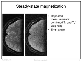
Steady-state diffusion TR< T1,T2
Navigated steady-state diffusion TR< T1,T2
Navigated phase correction • navigator measures motion-induced phase • phase-correct high resolution data (Poster #1112) kx kx ky ky high resolution navigator
In vivo experiments • 5 subjects • FOV = 18 cm, 0.75 x 0.75 mm2, 5 mm thick • TR = 25 ms, = 25, nex = 64 • G = {1, 2.75, 4} G/cm, t = 5 ms beff = 40, 275, 500 s/mm2 imaging time: 7 minutes
In vivo results 40 s/mm2 275 s/mm2 500 s/mm2
Navigator correction navigator corrected uncorrected
Conclusions • diffusion has potential as contrast mechanism in cartilage • SS-DWI enables in vivo knee cartilage diffusion imaging • navigated motion-correction allows high resolution