Download
1 / 41
430 likes | 751 Views
Diffusion Tensor Imaging. Johansen-Berg and Rushworth 2008 Glasser and Rilling 2008 20 April 2010. Connectivity. Connectivity patterns define functional networks Passingham et al. (2002) Connectional anatomy Boiling in oil Dissection Myelin stains Axonal stains

E N D
Diffusion Tensor Imaging Johansen-Berg and Rushworth 2008 Glasser and Rilling 2008 20 April 2010
Connectivity • Connectivity patterns define functional networks • Passingham et al. (2002) • Connectional anatomy • Boiling in oil • Dissection • Myelin stains • Axonal stains • Retrograde and anterograde tracers
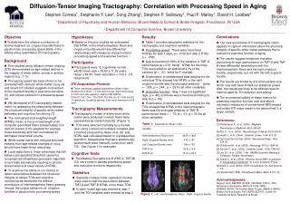
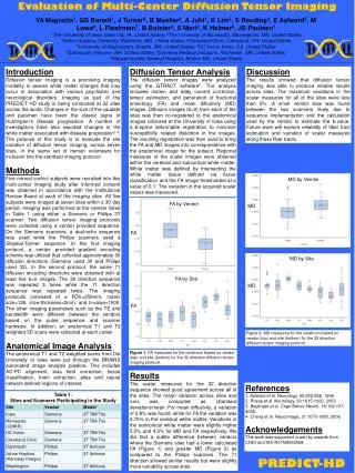
Diffusion-weighted MRI • Acquisition of multiple images • Each image is sensitized to a different direction so multiple measures are associated with each voxel • Fit a mathematical model such as diffusion tensor imaging • Each voxel is described by an ellipsoid, or tensor • Three orthogonal eigenvectors and associated eigenvalues (λ1, λ2, λ3)
Diffusion-weighted MRI • Self-diffusion of water molecules • Water is directionally dependent in tissue with directional structure
Fractional Anisotropy • In the ventricles, diffusion should be similar in each direction which is associated with a spherical tensor • Fiber bundles have more barriers and the associated tensor is elongated • Directional dependency (Anisotropy) is quantified by fractional anisotropy • 0 (fully isotropic) to 1 (fully anisotropic) • White matter: ~0.6-0.8
Diffusion Anisotropy • Directional estimates of principal diffusion direction form the basis of diffusion tractography • This is NOT anatomical • Functional connectivity is just the correlation between two or more active areas
Deterministic vs. Probabilistic Models • Difficult to interpolate beyond the end of the fiber bundle • One can assign a probability to the direction of each voxel • Sampling is used to create streamlines which are used to represent interconnectivity
Trace cortico-cortical networks Parietal and premotor cortex Fine-grained spatial mapping Thalamic connections Difficult to determine connection strength Anterograde or retrograde? Accurate detection of connections Validation: biologically realistic phantoms Strengths and Weaknesses
DTI Tractography of the Human Brain’s Language Pathways Glasser and Rilling 2008 http://cercor.oxfordjournals.org/cgi/content/full/bhn011/DC1
DTI and Language • Arcuate Fasciculus • Originates in temporal lobe • Curves around Sylvian fissure • Projects into the frontal lobe • Connects Broca’s area and Wernicke’s area • Dejerine 1895 • Leftward asymmetry in the arcuate fasciculus
Phonemes Wernicke’s area and BA 40 Posterior Broca’s area Bilateral Prosody Right MTG Frontal lobe Bilateral? Lexical-semantics Concepts and meanings Middle and temporal areas (BA 21 and 17) Broca’s area and areas more anterior and frontal Left localized Three Components of Langauge
Hypotheses • Bilateral arcuate connection to the superior temporal gyrus • Leftward asymmetry connection to middle temporal gyrus
Methodology • No behavioral tasks • A priori reasons to think phonology, lexico-semantics, and prosody have distinct locations • Determine the connections using a deterministic algorithm • Corroborate the connections using outside sources
Subjects • 20 right-handed males • 18-50 (23.75, 7.1) • Handedness and sex influence laterality
Diffusion MRI TE = 90 ms TR = 7700 ms 12 diffusion directions 1.7 x 1.7 x 2.0 mm voxels T1-weighted TE = 4 ms TR = 2300 ms 256 x 256 matrix MRI
Deterministic Tractography • Siemens DTI Task Card Parameters • # of samples/voxel length = 8 • Minimum FA threshold = 0.15 • Maximum turning angle = 15° • Step length between calculations = 0.25 mm
Deterministic Tractography • Step 1: • Select Arcuate Fasciculus as ROI in left and right hemispheres from a coronal slice • ROIs defined as BA 22 (Wernicke’s), BA 21 and BA 37 (middle temporal gyrus) • Determine terminations in frontal lobe ROI • BA 44, 45, 6, 9 (Broca’s area and surrounding cortex)
Left Hemisphere Found in 17 of 20 subjects BA 22 (posterior STG) connected to BA 44 and BA 6 Right Hemisphere Found in 4 of 20 subjects BA 22 (posterior STG) connected to BA 44 and BA 6 The STG Pathway
Left Hemisphere Found in 20 of 20 BA 21 and BA 37 projected to BA 44 and parts of BA 6, 9, 45 Right Hemisphere Found in 11 of 20 BA 37 (posterior MTG) The MTG Pathway
Left Hemisphere Phonological Studies overlapped with STG pathway Lexical-Semantic Studies overlapped with MTG, ITG, and AG segments Right Hemisphere Phonological Studies did not overlap (more anterior) with STG pathway Prosody Studies overlapped MTG and STG segments Functional Activations
Asymmetries in Connections • Phonology is usually bilaterally activated; more activation found on the left side than usual • Lexical-Semantics is usually activated on the left; more activation found on the right than usual
Aphasia • Can we explain aphasia using the Hickok and Poeppel (2004) and Price (2000) model with additional information added from the DTI study?
Broca’s Aphasia • Symptoms • Difficulty producing grammatical speech • Slow and halting speech • Able to communicate • Difficulty with phoneme discrimination • Location of lesions • Left inferior and premotor cortex (Dysarthria) • BA 44 and BA 45 (agrammatism) • Frontal cortical terminations
Wernicke’s Aphasia • Symptoms • Fluent speech • Frequent phonological and semantic errors • Difficulty understanding speech • Location of lesions • Left STG or subcortical connections • Middle and inferior temporal cortex • Disruption of phonological decoding
Conduction Aphasia • Symptoms • Difficulty with repetition • Phonological errors • Difficulty naming objects • Intact comprehension • Spontaneous speech • Location of lesions • Left supramarginal gyrus (BA 40) • Arcuate fasciculus? • Superficial to avoid damaging the MTG pathway
Conduction Aphasia • Symptoms • Difficulty with repetition • Difficulty naming objects • Phonological errors • Intact comprehension • Spontaneous speech preserved • Location of lesions • Left supramarginal gyrus (BA 40) • Superficial arcuate fasciculus
Transcortical Motor Aphasia • Symptoms • Limited spontaneous speech • Impaired naming • Intact repetition • Normal articulation • Good auditory comprehension • Location of lesions • Anterior and superior to Broca’s area • White matter near MTG pathway
Transcortical Sensory Aphasia • Symptoms • Intact repetition • Lack of understanding • Location of lesions • Left temporal lobe lesion which spares Wernicke’s area • Lesions in middle and inferior temporal lobe • Electrical stimulation of LMTG and LITG causes transient aphasia
Aprosodias • Symptoms • Difficulty with comprehension of emotional prosody • Location of lesions • Right temperoparietal lesions • Right middle and temporal cortex • Subcortical
Limitations • Unidentified pathways probably exist • Crossing fibers add noise • Deterministic vs. Probabilistic • Heterogeneity of functional areas • Only three components of language
Unexpectations • Lexical-semantic and STG pathways project to BA 6 • Fewer projections to BA 45 and BA 47 using a deterministic methodology than a probabilistic methodology • More anterior with probabilistic • These differences could be explained if the arcuate fasciculus was combined with the SLF
Conclusions • Arcuate fasciculus connects the STG and MTG with the inferior frontal lobe • Phonological processing associated with left STG • Lexical-semantic processing associated with left MTG • Prosody processing associated with right MTG and STG
Questions • Marianna • How can they find evidence of activation of different areas? • Lucy • What would these models predict for people who are relatively more bilateral? Just more/higher incidence of pathways in the right hemisphere?
Questions • Israel • How do they know that the connections they find are related to these linguistic areas and not to the other linguistic areas? • Pawel • Couldn't familial left-handedness explain the results showing that they were unable to find MTG pathways in the right hemisphere of some subjects and the STG pathway in the left hemisphere of others?
Questions • Lynn • Could the motor theory or elements of it be useful in interpreting any of the current results including visual information and the right hemisphere?
Neat Website • http://white.stanford.edu/~brian/papers/mri/2006-Wandell-NIPS-Tutorial.pdf