Download
1 / 20
200 likes | 392 Views
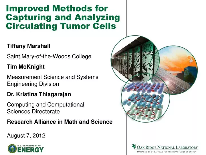
Improved Methods for Capturing and Analyzing Circulating Tumor Cells . Tiffany Marshall Saint Mary-of-the-Woods College Tim McKnight Measurement Science and Systems Engineering Division Dr. Kristina Thiagarajan Computing and Computational Sciences Directorate

E N D
Improved Methods for Capturing and Analyzing Circulating Tumor Cells Tiffany Marshall Saint Mary-of-the-Woods College Tim McKnight Measurement Science and SystemsEngineering Division Dr. Kristina Thiagarajan Computing and ComputationalSciences Directorate Research Alliance in Math and Science August 7, 2012
Outline Introduction Methods Results Future work Conclusion Questions
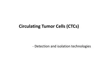
Circulating tumor cells (CTCs) • Found in bloodstream • Important to isolate and enrich • Facilitates early detection in malignant disease patients • More effective treatment
Commercial systems for CTCenrichment • CTCs are rare in bloodstream • Magnetic beads used to capture CTCs from a vastmajority of non-CTC cells • Bead capture is harsh and results in phenotypic disturbances or loss of viability Veridex CellSearch system is used to isolate CTCs
Emerging microfluidic systemsfor CTC enrichment • Requires large processing volumes (7ml of blood) • Low flow throughout requires long processing times • Shear stresses still cause phenotypic shift or even loss of viability From Stott et al (2010) PNAS Demonstrated effective, but with very limited throughput
ORNL designed microfluidiccapture and analysis platform • Vertically-aligned carbon nanofibers (VACNF) • Cellular isolation • Direct gene delivery • Fabricated at the Center for Nanophase Materials Sciences (CNMS) McKnight et al, CRC Nanotechnology in Biology and Medicine, 2007.
ORNL methods should improve capture efficiency and cell viabilityenabling post enrichment assay • Maintain cell viability due to lower shear stress • Higher throughput (due to smaller flow restriction) • Faster processing times vs.
Development of post enrichmentlive cell assay methods • Cell culture • Plasmid preparation • Rapid transgene assay • Analysis of transgene expression
Cell culture • MCF-7 cell line used as a surrogate of metastatic breast cancer • Cultured in Dulbecco’s Modified Eagles Medium (DMEM) • 10% fetal bovine serum • Penicillin • Non-essential amino acids • L-glutamine • Sodium pyruvate • Feed cells every 2-3 days • Aspirate old media • Add 5 new mls • Harvest cells approximately twice a week • Experimental use • Room to grow • Place in incubator to grow with CO2 environment
Plasmid preparation • Use pd2eYFP-N1 plasmid that encodes destabilized yellow fluorescent protein (YFP) used as a marker for rapid transgene expression • Heat shock plasmid into competent e. coli bacterial cells • Transformed bacteria resist antibiotic selection and produce high copies of plasmid DNA • Grow bacterial cultures overnight • Chemically lyse cultures so high copy DNA plasmid can be extracted and purified using affinity-based kits.
Rapid transgene assay • Prepared VACNF chips using ambient air plasma • Applied 100 ng of yellow fluorescent protein plasmid (pd2eYFP) to chip and dried • Centrifuged cells into pellet • Impaled pelleted cells onto chip • Used time lapse fluorescence microscopy to observe onset of transgene expression
Analysis of transgene expression • Impaled cells receiving DNA are tracked usingtimelapse fluorescence microscopy • Acquired images 1/min at 1500 msec exposure • Analyzed and processed image data • Used image J software • MatLab routine todeterminefluorescencetrajectories anddata binning
Results • Maintain cell viability following rapid transgene technique • YFP rapidly expressed and observed • Earliest transgene expression observed within 50 min. • Average transgene expression observed ~ 130 min. • Timeframes are consistent with nuclear delivery
Next step - delivery of TRF2-GFP transgene • Telomere binding protein (TRF2) mislocalizes in metastatic breast cancer cells • Delivery and expression of TRF2-GFP provides visualization of TRF2 localization within cells • Rapid transgene studies • Assess metastatic potential of cells • In-vitro studies of cellular response to chemotherapy
Fluorescence in situ hybridization (FISH) probing • Nuclear delivery of fluorescent probe • Use to measure HER2 gene amplification • Test positive for HER2 amplification • Higher mortality rate • Higher rate of cancer reoccurring
Conclusion • Deliver genes into nucleus of MCF-7 cells is possible while maintaining viability • Plasmid DNA delivery results in rapid transgene expression • Nuclear delivery of probes • Continued viability of interfaced MCF-7 cells • Establishes rapid transgene assay as a potential means of tailoring therapeutic intervention using enriched CTCs from clinical samples
Acknowledgments • A special thanks to Tim McKnight, Morten Madsen, and Dr. Kristina Thiagarajan for their mentoring. • The Research Alliance in Math and Science program is sponsored by the Office of Advanced Scientific Computing Research, U.S. Department of Energy. • The work was performed at the Oak Ridge National Laboratory, which is managed by UT-Battelle, LLC under Contract No. De-AC05-00OR22725. This work has been authored by a contractor of the U.S. Government, accordingly, the U.S. Government retains a non-exclusive, royalty-free license to publish or reproduce the published form of this contribution, or allow others to do so, for U.S. Government purposes.
Sources • Melechko, Anatoli V, R. D. (2009). Synthesis of vertically aligned carbon nanofibres for interfacing with live systems. Journal of Physics D: Applied Physics . • How many women get breast cancer? (2012, March 12). Retrieved June 1, 2012, from American Cancer Society: http://www.cancer.org/Cancer/BreastCancer/OverviewGuide/breast-cancer-overview-key-statistics • Lui, M., Griffin, G., & McKnight, T. (n.d.). Hybrid Nanostructured/Microstructured Platforms for the Enrichment of Circulating Tumor Cells. Seed Proposal to the Georgetown/ORNL CTSA . • Markman, Maurie B. (2012, June 13). Breast Cancer and HER2 Overview of HER2 Breast Cancer. Retrieved July 12, 2012, from Medscape Reference: http://emedicine.medscape.com/article/1689966-overview#aw2aab6b4 • Liu, Minetta C., P. G. (2009). Circulating Tumor Cells: A Useful Predictor of Treatment. Journal of Clinical Oncology , 5153-5158. • Nijjar, T. (2004). Accumulation and altered localization of telomere-associated protein TRF2 in immortally. Lawrence Berkeley National Laboratory . • Stott, Shannon L. b. C.-H. (2010). Isolation of circulating tumor cells using a microvortex-generating herringbone-chip. PNAS , 18392-18397. • Nagrath, Sunitha L. V. (2007). Isolation of rare circulating tumour cells in cancer patients by microchip technology. Nature , 1235-1238.