Download
1 / 60
600 likes | 814 Views
Renal Failure in the ED. CCFP(EM) Core Lecture Presentation University of Ottawa Dr. Debra J. Weatherhead, MD, CCFP (EM), FCFP April 25, 2013. Learning Objectives. 1) Know the terminology associated with renal failure. 2) Know the causes and ED management of Acute Renal Failure (ARF).

E N D
Renal Failure in the ED CCFP(EM) Core Lecture Presentation University of Ottawa Dr. Debra J. Weatherhead, MD, CCFP (EM), FCFP April 25, 2013
Learning Objectives 1) Know the terminology associated with renal failure. 2) Know the causes and ED management of Acute Renal Failure (ARF). 3) Know the ED management of dialysis related emergencies.
Renal failure ..... Who?
Older Diabetic Hypertensive
A Case in Review • 8 yo F. Previously healthy. • Seen by FD #1 Wed for watery D. • F/U arranged for Fri afternoon. • Fri afternoon: drinking but still D. Since the am, blood in D. Dehydrated. Arrangements made for admission, rehydration & investigations.
Arrived at hosp @ 6pm. • Bloodwork done. • Stool cultures sent. • Rehydration started .
Sat am: FD # 2 doing rounds notes abN lytes , elevated WBC & sl decreased Plts. • Peds #1 consulted. • Dx SIADH. Fluid restricted. • Febrile. Bloody D & anorexia persisting.
Sun am: FD # 3 morning rounds & Peds #1. Fluid restriction continued. • Sun pm FD #3 called re: increased abdo pain --> Demerol 60 mg IM ordered. • Mon 0100 hr FD #3 called re: increased abdo pain--> Demerol 60 mg IM ordered
Mon 0500 hr FD 3# called: much worse, increased abdo pain, tachycardiac, hallucinating, worsening dehydration …Demerol 60mg IM ordered.
0720 hr Mon am: FD #1 signs into hospital computer --> immediately receives stat call to peds.
Looks ill. Looks toxic. Child is SICK !!! • Tachycardiac. Tachypneic. Febrile. • Severely dehydrated. Hallucinating. • Moaning in pain - lying rigidly. • Bed bumped -- child screams in pain.
rigid abdo • marked signs of peritonitis • multiple areas of petechaie & purpura
Markedly elevated WBC • Decreased Hgb • Markedly decreased Plts • Creat > 1300 BUN ~ 30 • Hyperkalemic
Stool cultures ………….. • …….. E . Coli 0157H7
Anticipated lifetime anti - HTNs. • Amazingly, still N cognitive status but felt that she did lose ~ 5% of her previous cognitive functioning.
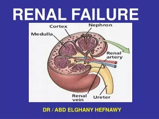
Terminology • Uremia = contamination of the blood with urine • Azotemia = accumulation of nitrogen in the blood • Urea = the major breakdown product of proteins • Creatinine = a breakdown product of the skeletal muscle protein creatine
The normal functions of the kidneys are: • glomerular filtration • tubular reabsorption • secretion
Acute Renal Failure (ARF) • The deterioration of renal function over hrs/days accumulation toxins loss of homeostasis.
Case 1 • 38 yo healthy male • onset watery diarrhea 1200hrs • “too sick” to try po fluids • diarrhea x at least 40 times • you see Px at MN
HR-160 80/50 • RR-not done 94% R/A • conscious, looks ashen • states “too weak to talk” • manage this patient….
Causes ofAcute Renal Failure (ARF) • Prerenal – decreased perfusion of N kidney - glomerular & tubular fn intact • Renal/Intrinsic – pathology of the kidney • - dz of glomerulus, interstitium, small vessels, tubules • assoc with release of renal vasoconstrictors • most common cause is ischemic ARF (ATN) = acute kidney injury • Postrenal – obstruction of urinary outflow
Community-acquired ARF • –most common cause is prerenal • - most common prerenal cause is volume depletion Hospital-acquired ARF - most common cause is renal/intrinsic
DDx of Prerenal ARF • Hypovolemia • Hypotension (frank and relative) • Renal artery & small vessel disease • emboli, thrombosis, microvasc thrombosis (DIC, HUS, vasculitis, sickle cell, preeclampsia), medications
DDx of Intrinsic ARF • tubular disease • ischemia, nephrotoxins (meds, IV contrast, heme pigments) • interstitial disease • interstitial nephritis (most commonly 2’ meds), infection, sarcoidosis, autoimmune (SLE) • glomerular disease • Goodpasture syndrome, Wegener, HSP • small vessel disease • microvasc thrombosis (DIC, HUS,TTP,vasculitis), malignant HTN, scleroderma
DDx of Postrenal ARF • Ureter – stone, stricture, tumor • Urethra & bladder outlet – BPH, Ca, phimosis, obstructed catheter, neurogenic bladder, clot, trauma • Peds – anatomic malformations
In postrenal ARF, if complete obstruction, significant permanent loss of renal function after 10-14 days. • Risk of permanent renal failure increases significantly if obstruction is complicated by UTI. • A postvoid bladder residual > 125 cc suggests obstruction ..... Keep catheter in until obstruction resolved.
Case #2 • 45 yo healthy male • Norwalk outbreak • 36 hrs V + D • to ED via EMS • CTAS 1 to resusc
EMS unable to get bp • O2sat not registering • sinus brady (30-32 bpm) • Px obtunded, incoherent, occasional moaning • Manage this patient ……
RIFLE Classification of ARF GFRUrine output Risk serum Cr x 1.5 0.5cc/kg/h x 6h Injury serum Cr x 2.0 0.5cc/kg/h x 12h Failure serum Cr x 3.0 0.3cc/kg/h x 24h or anuria x 12h Loss complete loss of kidney fn > 4 wks ESRD need renal replacement for> 3 mo
Imaging in ARF • U/S is imaging of choice • non-contrast CT also a consideration
Management of ARF • * • * • * • * • *
Stages of Chronic Renal Disease StageDescriptionGFR 1 kidney damage (protein in urine) >90 & normal GFR 2 kidney damage & mild decrease GFR 60-89 3 moderate decrease in GFR 30-59 4 severe decrease in GFR 15-29 5 kidney failure (dialysis/transplant) < 15
ESRD • Uremia • - contamination of the blood with urine • - the clinical syndrome resulting from ESRD
Pathophysiology of Uremia • Excretory failure – leads to build up of chemicals in blood • Biosynthetic failure – kidneys produce erythropoietin & 1alpha hydroxylase (needed for Vit D - resulting in anemia & renal bone disease • Regulatory failure – results in over secretion of hormones
Case # 3 • 64 yo male Hemodialysis • Intentionally missed last 2 dialysis treatments. • To ED via EMS – presyncopal, diaphoretic, gray, looks unwell • Manage this patient…..
Most Common Reasons for Emergent Dialysis • Hyperkalemia • Severe acid-base disturbances • Pulmonary edema resistant to other Mx
Dialysis • Hemodialysis • ~ 4 hrs per day every other day • Peritoneal Dialysis • Uses 4 exchanges daily (4 x 2 litres) with 10 litres off.
Case # 4 • 28 yo male Peritoneal dialysis • 2-3 days Hx feeling unwell. Today fevers, chills. • T- 38.9 HR – 114 RR- 16 142/76 98%r/a • Manage this patient……
Peritonitis • Most common complication of PD. • Fever, adbo pain, rebound tenderness • Cloudy effluent • Cell count > 100 leukocytes (>50% neutrophils) • Gram stain, + only 10-40% cases of culture + peritonitis • Expect gram positive & gram negative
Empiric AB coverage in dialysate • 1st gen cephalosporin 500mg/L 1st exchange then 200mg/L subsequent exchanges. • Vancomycin 500mg/L 1st exchange then 50mg/L subsequent exchanges. • Gentamycin 100mg/L 1st exchange then 4-8mg/L subsequent exchanges.
Case # 5 • 56 yo male • R flank pain, dysuria, polyuria, R testicular pain, microhematuria • diagnosis?
CT 9mm proximal ureteral stone • WBC – 7.5 • Creat – 100 • BUN – 5.6 • U/A – trace RBC, all else neg
Returns to ED 3 days later. • Fevers, chills, feels unwell. • WBC- 16.9 (with left shift) • Creat – 529 • BUN – 12.4
Case # 6 • 5 yo healthy female. • WIC for fever and ST Dx’ed strep throat Amoxil • 5 days later local community ED for V +++ • - told bp high but probably due to V • - advised continue Amoxil • 2 days later WIC gross hematuria and persistent V
Case # 7 • 65 yo female HTN, DM (controlled) • Seen in ED by PGY 3 (non-cardiac CP) • Incidentally, creat – 130 • D/C’ed home