Download
1 / 50
540 likes | 913 Views
Renal Failure. Prepared by Miss Fatima Hirzallah. What is renal failure?. renal failure. Renal failure refers to temporary or permanent damage to the kidneys that results, when the kidneys cannot remove the body's metabolic wastes or perform their regulatory functions.

E N D
Renal Failure Prepared by Miss Fatima Hirzallah
renal failure • Renal failure refers to temporary or permanent damage to the kidneys that results, when the kidneys cannot remove the body's metabolic wastes or perform their regulatory functions. • The substances normally eliminated in the urine accumulate in the body fluids as a result of impaired renal excretion ,leading to a disruption in endocrine and metabolic functions as well as fluid, electrolyte, and acid–base disturbances .
There are two different types of renal failure -acute renal failure (ARF) - chronic renal failure (ESRD)
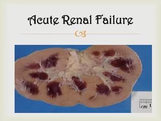
Acute renal failure • Acute renal failure (ARF) is a reversible clinical syndrome where there is a sudden and almost complete loss of kidney function (decreased GFR) over a period of hours to days with failure to excrete nitrogenous waste products and to maintain fluid and electrolyte homeostasis
The hallmark of acute renal failure is • a decreased glomerular filtration rate (GFR), reflected by an accumulation of blood urea nitrogen (BUN) and serum creatinine- a condition termed azotemia . • Serum creatinine is the better marker because increases in serum creatinine are relatively unaffected by non -renal mechanisms.
Urine volume may be normal, or changes may occur. Possible changes include • oliguria (less than 400 mL/day), • nonoliguria (greater than 400 mL/day), • or anuria (less than 50 mL/day)
Pathophysiology • Although the exact pathogenesis of ARF and oliguria is not always known, many times there is a specific underlying problem . • the following conditions that reduce blood flow to the kidney and impair kidney function: • (1) hypovolemia; (2) hypotension; (3) reduced cardiac output and heart failure; (4) obstruction of the kidney or lower urinary tract by tumor, blood clot, or kidney stone; and (5) bilateral obstruction of the renal arteries or veins .
If these conditions are treated and corrected before the kidneys are permanently damaged, the increased BUN and creatinine levels, oliguria, and other signs associated with ARF may be reversed.
Categories of Acute Renal Failure • The major categories of ARF are • prerenal (hypoperfusion of kidney), • intrarenal (actual damage to kidney tissue), and postrenal (obstruction to urine flow).
Causes of Acute Renal Failure • Prerenal ARF, which occurs in 60% to 70% of cases, is the result of impaired blood flow that leads to hypoperfusion of the kidney and a decrease in the GFRVolume depletion resulting from: • Hemorrhage • Renal losses (diuretics, osmotic diuresis) • Gastrointestinal losses (vomiting, diarrhea, nasogastric suction)
Impaired cardiac efficiency resulting from: • Myocardial infarction • Heart failure • Dysrhythmias • Cardiogenic shoc • Vasodilation resulting from: • Sepsis • Anaphylaxis
Intrarenal ARF is the result of actual parenchymal damage to the glomeruli or kidney tubules. Nephrotoxic agents, such as aminoglycosides and radiocontrast agents, account for 30% of cases of acute tubular necrosis (ATN), and ischemia due to decreased renal perfusion accounts for more than 50% of cases of ATN.
Intrarenal Failure • Prolonged renal ischemia resulting from: • Pigment nephropathy (associated with the break-down of blood cells containing pigments that in turn occlude kidney structures) • Myoglobinuria (trauma, crush injuries, burns) • Hemoglobinuria (transfusion reaction, hemolytic anemia)
Infectious processes such as: • Acute pyelonephritis • Acute glomerulonephritis • Nephrotoxic agents such as: • Aminoglycoside antibiotics (gentamicin, tobramycin) • Radiopaque contrast agents • Solvents and chemicals (ethylene glycol, carbon tetrachloride, arsenic) • Nonsteroidal anti-inflammatory drugs (NSAIDs) • Angiotensin-converting enzyme inhibitors (ACE inhibitors)
Postrenal ARF is usually the result of an obstruction somewhere distal to the kidney. Pressure rises in the kidney tubules and eventually, the GFR decreases. • Urinary tract obstruction, including: • Calculi (stones) • Tumors • Benign prostatic hyperplasia • Strictures • Blood clots
Phases of Acute Renal Failure • There are four clinical phases of ARF: initiation, oliguria, diuresis, and recovery
The initiation period begins with the initial insult and ends when oliguria develops. • The oliguria period is accompanied by an increase in the serum concentration of substances usually excreted by the kidneys (urea, creatinine, uric acid, organic acids, and the intracellular cations [potassium and magnesium]).
The diuresis period is marked by a gradual increase in urine output, which signals that glomerular filtration has started to recover. Laboratory values stop increasing and eventually decrease. Although the volume of urinary output may reach normal or elevated levels, renal function may still be markedly abnormal .
The recovery period signals the improvement of renal function and may take 3 to 12 months. Laboratory values return to the patient's normal level. Although a permanent 1% to 3% reduction in the GFR is common, it is not clinically significant .
Clinical Manifestations • The patient may appear critically ill and lethargic. The skin and mucous membranes are dry from dehydration. Central nervous system signs and symptoms include drowsiness, headache, muscle twitching, and seizures.
Assessment and Diagnostic Findings • In ARF, urine output varies (scanty to normal volume), hematuria may be present, and the urine has a low specific gravity (compared with a normal value of 1.003 to 1.030). One of the earliest manifestations of tubular damage is the inability to concentrate the urine
with prerenalazotemia have a decreased amount of sodium in the urine (less than 20 mEq/L) and normal urinary sediment. Patients with intrarenalazotemia usually have urinary sodium levels greater than 40 mEq/L with urinary casts and other cellular debris. • Ultrasonography is a critical component of the evaluation of patients with renal failure. A renal sonogram or a CT or MRI scan may show evidence of anatomical changes
The BUN level increases steadily at a rate dependent on the degree of catabolism (breakdown of protein), renal perfusion, and protein intake. Serum creatinine increases in conjunction with glomerular damage. Serum creatinine levels are useful in monitoring kidney function and disease progression . hyperkalemia (high serum potassium levels).
Prevention • Management of ARF is expensive and complex, and even when optimal, the mortality rate can be as high as 60% to 80%, depending on etiology and comorbidities Therefore, prevention of ARF is essential
A careful history is obtained to determine whether the patient has been taking potentially nephrotoxic antibiotic agents or has been exposed to environmental toxins
Medical Management • The objectives of treatment of ARF are to restore normal chemical balance and prevent complications until repair of renal tissue and restoration of renal function can occur. Management includes maintaining fluid balance, avoiding fluid excesses, or possibly performing dialysis. • The underlying cause is identified, treated, and eliminated when possible
Pharmacologic Therapy • Hyperkalemia is the most life-threatening of the fluid and electrolyte changes that occur in patients with renal disturbances • Therefore, the patient is monitored for hyperkalemia through serial serum electrolyte levels (potassium value more than 5.5 mEq/L [5.5 mmol/L]), ECG changes (tall, tented, or peaked T waves), and changes in clinical status.
The elevated potassium levels may be reduced by administering cation-exchange resins (sodium polystyrene sulfonate [Kayexalate]) orally or by retention enema. Kayexalate works by exchanging sodium ions for potassium ions in the intestinal tract.
Many medications are eliminated through the kidneys; therefore, medication dosages must be reduced when a patient has ARF. • Examples of commonly used agents that require adjustment are antibiotic medications (especially aminoglycosides), digoxin, ACE inhibitors, and magnesium-containing agents
In patients with severe acidosis, the arterial blood gases and serum bicarbonate levels must be monitored because the patient may require sodium bicarbonate therapy or dialysis. If respiratory problems develop, appropriate ventilatory measures must be instituted. The elevated serum phosphate level may be controlled with phosphate-binding agents (aluminum hydroxide).
Nutritional Therapy • ARF causes severe nutritional imbalances (because nausea and vomiting contribute to inadequate dietary intake), impaired glucose use and protein synthesis, and increased tissue catabolism. • Dietary proteins are individualized to provide the maximum benefit
(Caloric requirements are met with high-carbohydrate meals, because carbohydrates have a protein-sparing effect (ie, in a high-carbohydrate diet, protein is not used for meeting energy requirements but is “spared” for growth and tissue healing).
The oliguric phase of ARF may last 10 to 20 days and is followed by the diuretic phase, at which time urine output begins to increase, signaling that kidney function is returning. Results of blood chemistry tests are used to determine the amounts of sodium, potassium, and water needed for replacement, along with assessment for overhydration or underhydration.
Following the diuretic phase, the patient is placed on a high-protein, high-calorie diet and is encouraged to resume activities gradually
Foods and fluids containing potassium or phosphorus (eg, bananas, citrus fruits and juices, coffee) are restricted. Potassium intake may be restricted, and the patient may require parenteral nutrition.
Nursing Management • Monitoring Fluid and Electrolyte Balance • Reducing Metabolic Rate • Promoting Pulmonary Function • Preventing Infection • Providing Skin Care • Providing Support
What is dialysis? • Dialysis is a procedure that is performed routinely on persons who suffer from acute or chronic renal failure, or who have ESRD. • The process involves removing waste substances and fluid from the blood that are normally eliminated by the kidneys. • Dialysis may also be used for individuals who have been exposed to or ingested toxic substances to prevent renal failure from occurring.
dialysis • There are two types of dialysis that may be performed, including the following: • peritoneal dialysis • hemodialysis
Indications for Hemodialysis • Hemodialysis is indicated in chronic renal failure and for complications of acute renal failure. These include uremia, fluid overload, acidosis, hyperkalemia, and drug overdose.
Contraindications to Hemodialysis • Hemodialysis may be contraindicated in patients with coagulopathies because the extracorporeal circuit needs to be heparinized. • Hemodialysis may also be difficult to perform in patients who have extremely low cardiac output or who are sensitive to abrupt changes in volume status.
Methods of vascular access for hemodialysis. (A) Arteriovenous fistula. (B) Synthetic graft.
hemodialysis • Hemodialysis is can be performed at home or in a dialysis center or hospital by trained healthcare professionals. • A special type of access, called an arteriovenous (AV) fistula, is placed surgically, usually, in the arm. This involves joining an artery and a vein together.
An external, centralvenous catheter (CVC) may also be inserted, but is less common for long-term dialysis. • After access has been established, you will be connected to a large hemodialysis machine which drains the blood, bathes it in a special dialysate solution which removes waste substances and fluid, then returns it to your bloodstream .
hemodialysis • An AV fistula requires advance planning because a fistula takes a while after surgery to develop. But a properly formed fistula is less likely than other kinds of vascular access to form clots or become infected. Also, properly formed fistulas tend to last many years—longer than any other kind of vascular access
arteriovenous graft • What is an arteriovenous graft? • If you have small veins that won’t develop properly into a fistula, you can get a vascular access that connects an artery to a vein using a synthetic tube, or graft, implanted under the skin in the arm. The graft becomes an artificial vein that can be used repeatedly for needle placement and blood access during hemodialysis.
Compared with properly formed fistulas, grafts tend to have more problems with clotting and infection and need replacement sooner. However, a well-cared-for graft can last several years.
Equipment • DIALYZERS hemodialysisThe hollow-fiber dialyzer is the most commonly used configuration. In this design, the blood path flows through hollow fibers composed of semipermeable membrane, and the dialysate path is encased in a rigid plastic tube. Dialysate surrounds each hollow fiber. This provides a large surface area to cleanse the blood.
Blood and dialysate flow in opposite directions from each other (countercurrent flow); as blood travels through the dialyzer, it is constantly exposed to a fresh flow of dialysate. • This countercurrent flow maintains the concentration gradient between the two compartments and provides the most efficient dialysis