Download
1 / 49
500 likes | 734 Views
Diagnosis and Treatment of Infection Following Total Hip Arthroplasty. ICL 34 & 35. Introduction. In the 1960’s, Charnley reported a rate of infection of 9.5%. More recently, authors have reported that infection causes failure after 1-2% of primary THAs.

E N D
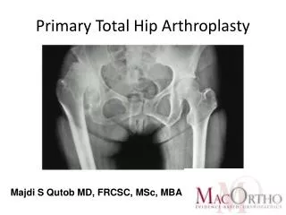
Diagnosis and Treatment of Infection Following Total Hip Arthroplasty ICL 34 & 35
Introduction • In the 1960’s, Charnley reported a rate of infection of 9.5%. • More recently, authors have reported that infection causes failure after 1-2% of primary THAs. • In the US, the cost per year to treat the 3,500-4,000 infections following THA is between 150 and 200 million dollars.
Introduction • Infection following THA can present a diagnostic challenge • No test is 100% sensitive and 100% specific. • The diagnosis then relies heavily upon the surgeon’s judgement of the clinical presentation, the findings on PE, and the interpretation of the results of previous investigations. Misdiagnosis can be critical.
Clinical Presentation • A thorough H&P are of paramount importance in the Dx of infection. • Coventry and later Fitzgerald and assoc. described the most common system for the classification of infection after THA. • Type-I • Type-II • Type-III
Type-I Infections • These occur immediately in the postop period--usually during the first month. • Systemic signs include: fever, chills, sweating, and continuous pain. • Wound appears erythematous, swollen, fluctuant, and tender, and if drainage is present it is usually purulent. • Caused by infected hematomas or superficial wounds spreading below the fascia.
Type-II Infections • These are also believed to begin at the time of surgery. • Usually seen between 6 and 24 months after procedure • Hallmark is a gradual deterioration in function and an increase in pain. • Often, the only clue to infection is early loosening of the components. • Systemic Sxs are not part of the presentation.
Type-III Infections • These are the least common and are caused by a hematogenous spread to a previously asymptomatic hip, usually 2 years or more after arthroplasty. • Generally, there is an acute febrile episode followed by a sudden , rapid deterioration of the hip. • Dx can usually be made on the basis of the H&P.
Type-III Infections • These are likely to occur in patients who are immunosuppressed, have recurrent bacteremia, such as IVDAs, or those who need repeat urinary catheterization. • Other factors with type-III are dental manipulation, respiratory infections, open skin lesions, and endoscopy.
Preoperative Investigations • White Blood-Cell Count • Erythrocyte Sedimentation Rate • C-Reactive Protein Level • Plain Radiography • Radionuclide imaging • Other Imaging Modalities • Hip Joint Aspiration • Molecular Analysis
White Blood-Cell Count • WBC is rarely abnormal in patients who have infection following THA--studies show approximately 15%. • If the patient does have an abnormal count, the systemic infection is usually clinically obvious and is either type-I or type-III.
Erythrocyte Sedimentation Rate and C-Reactive Protein Level • The ESR and the CRP level are the most useful screening labs for the diagnosis of a potential infection following THA. • RBCs have negative charges and acute phase reactants have positive charges. • An elevated ESR is an indirect indicator of an abundance of acute-phase reactants. • Values of > 30 or 35 mm /hour are generally considered to be abnormal.
C-Reative Protein Levels • Synthesized in the liver and is found only in trace amounts under normal conditions. • It rises in a nonspecific manner as a result of infectious, inflammatory, or neoplastic disorders. • It reaches its maximum values within 48 hours after surgery, then returns to trace amounts in aprox. 2-3 weeks. Therefore, it is a more sensitive indicator of infection.
Plain Radiography • Plain radiographs should be made for all patients who have a failed arthroplasty, even though they are of limited value as an investigative tool for the dx of infection. • Many radiographic findings are found in both septic and aseptic failure. • Periosteal new bone formation, with/without loosening of a component, has been considered pathognomonic of deep infection.
Plain Radiography • Early loosening and rapidly progressive radiolucent lines are also suggestive of infection. • Evidence of loosening of the femoral component involves radiolucency along the stem-cement interface, fracture of the cement mantle or the stem, or migration of the prosthesis.
Plain Radiography • Acetabular loosening is indicated by migration of the socket or the cement mantle, protrusio acetabuli, or acetabular fracture. • Athrography can improve the accuracy in diagnosing loosening especially on the acetabular side.
Radionuclide Imaging • Technetium-99m bone scans are sensitive but not specific. • A negative bone scan can rule out infection. • Many conditions can result in increased uptake for as long as one year after an uncomplicated THA and as long as 2 years after insertion of a prosthesis without cement.
Radionuclide Imaging • Gallium-67 citrate is an isotope that accumulates in areas of inflammation. • It is also non-specific as any process resulting in reactive bone formation may cause increased uptake. • Indium-111-labeled white blood cells are useful for the dx of conditions of increased vascularity and white blood-cell uptake. This has been used in combination with technetium and had higher sensitivities and specificities.
Radionuclide Imaging • Radiolabeled immunoglobulin-G has been used for the investigation of musculoskeletal infections. • One advantage is that the patient does not have to have phlebotomy before the scan is made. • Until additional studies are performed, the routine use of In-IgG scans cannot be recommended.
Radionuclide Imaging • Currently, the use of sequential Tc-99 and In-111-WBC scans is being recommended, however, the use of radiolabeled IgG may supersede the use of sequential scans provided they prove to be superior for the dx of infection in THA.
Other Imaging Modalities • MRI can be of value after an infection has been diagnosed . MRI can be used to determine the extent of the cement mantle within the femur and the pelvis so that the revision procedure can be planned appropriately. • Ultrasound can be used to measure the thickness of the joint capsule, which indicates infection. Also soft -tissue abscesses can be evaluated.
Hip Joint Aspiration • This is perhaps the most useful investigative tool for definitive confirmation of the presence or absence of infection. • Now most authors favor a more limited role, with aspiration being used to confirm a clinical suspicion of infection. • It can also support or negate the findings of other labs such as ESR and CRP that may be falsely elevated in connective-tissue dx.
Aspiration • An additional benefit is the ability to identify the organism and its antibiotic-sensitivity profile, which may influence preoperative planning. • The reported rates of sensitivity and specificity have varied widely. This suggests that aspiration is better for ruling infection in than for ruling it out. • All ABX should be discontinued for 2-3 weeks before the aspiration.
Aspiration • Local anesthetics should be used only for the skin and not in the joint as they are bacteriostatic. • Three samples are taken and if all three are positive a dx of infection is made.
Intraoperative Investigations • Intraoperative Frozen Sections--if ten or greater PMNs are found in the high-power field, this is evidence of a probable infection. • Gram Stain--these may be specific, but it lacks any acceptable level of sensitivity.
Intraoperative Cultures • Preoperative Abx should be withheld until specimens have been obtained • Clean instruments that have not been used on the skin should be used to obtain the specimens. • Samples should be taken close to the prosthesis and from inflamed tissue. • A minimum of three tissue specimens should be sent fresh for immediate processing.
Molecular Analysis • Molecular technology may be used to diagnose the presence of bacterial DNA and RNA. • Polymerase Chain Reaction (PCR) enables to production of large amounts of specific sequences of target DNA from small quantities of starting material. • It is susceptible to contamination because of its extremely high sensitivity to any bacterial particles.
Following a careful H&P, both the ESR and CRP level should be determined. If both results are normal and there is no suggestion of infection clinically, no additional investigations are needed. If the ESR or CRP level is elevated for any reason or there is clinical suspicion of infection, then an aspiration of the hip joint should be performed. Protocol for the DX of Infection
A dx is made if the clinical suspicion in high; the ESR or the CRP level, or both, are elevated for no other known reason; and the cultures of the aspirated fluid are positive. If the ESR or the CRP level, or both, are falsely elevated, an intraoperative frozen section may be used to confirm the dx. A sequential indium bone scan may be used preoperatively if the frozen section will not be available. Protocol--continued
The single most important factor in determining the treatment options for a patient in whom a THA has failed is the exclusion of a dx of infection. Conclusion
It is estimated that 200,000 THA will be performed this year in the US and that more than 4,000 new cases of periprosthetic hip infections will need treatment. There are considerable financial implications also involved in revision THA. A longer stay in the hospital, longer OR time, greater blood loss, higher rate of complications, as well as a higher cost of the implants. It is estimated that the cost of tx of an infected THA is $50,000. Treatment of Infection at the Site of THA
Debridement with retention of the prosthesis An immediate one-stage exchange arthroplasty An excision arthroplasty-either as a definitive, permanent procedure or as the first of a two or even three-stage reconstructive procedure. Surgical Treatment Options for Infected Total Hip Prosthesis
Antibiotics may be used as an adjunct to surgery either systemically or locally ( with the use of bone cement as the vehicle), or both. They may be used either to eradicate the infection or to chronically suppress the infection without surgical intervention. Antibiotic Usage
Staphylococcus aureus and Staphylococcus epidermidis are the most common infecting organisms. These are followed by a wide range of gram- positive and gram negative bacteria. More than 95% of the S aureus are sensitive to oxacillin and therefore a cephalosporin, however, S epidermidis has up to a 30% resistance to oxacillin. Microbiologic Considerations
Recent attention has been focused on the ability of an infecting organism to produce a slime layer, or glycocalyx. This layer takes time to form. Bacteria that exist within this biofilm are at least 500 times more resistant to antibiotics than the planktonic forms. They are also resistant to complement activation and neutrophil injestion. Micro Considerations
1. Old healed incisions should be used to gain access to the hip provided that the surgical exposure is not compromised. 2. Antibiotics should be withheld until the hip-joint capsule has been incised and specimens have been obtained. Surgical Considerations in Revisions
3. The choice of surgical approach should be based on the need to remove all foreign material and dead tissue, including bone, while at the same time avoiding devascularization of the tissue. 4. When all necrotic tissue and foreign material have been removed, the wound should be copiously irrigated with saline solution.
Antibiotics Without Surgery Debridement with Retention of the Prosthesis Girdlestone Arthroplasty Single-Stage Exchange Arthroplasty Two-Stage Exchange Arthroplasty Treatment Protocols
Most commonly used in the form of suppressive therapy when the patient is unfit to undergo major surgery or simply refuses further surgical treatment. The infecting organism must be known as well as the sensitivity and MIC. The antibiotic must be well tolerated by the patient or noncompliance will result. Antibiotics without Surgery
There is little argument about the necessity to remove a loose prosthesis from a chronically infected joint, however, removal of a well-fixed total hip implant carries the risk of causing major damage to the remaining bone stock. Tsukayama and associates emphasized the importance of limiting this tx to infections that developed less than 1 month postop. Debridement with Retention of the Prosthesis
This did not allow the organisms to produce the resistant slime layer and could therefore be controlled. The primary difficulty appears to be the lack of accuracy with which acute infections can be distinguished from chronic ones. This procedure can only be implemented if the history of the infection can be accurately identified.
The general consensus is that the procedure is highly effective in controlling infection and reducing pain; however, it usually is associated with a considerable loss of function. Patients walk poorly and almost always need walking aids. Limb shortening may range from 3-11cm but most typically ranges from 4-6 cm. Girdlestone Arthroplasty
This may be appropriate for patients who are mentally impaired and who are unable to cooperate with the postoperative restrictions. Excision arthroplasty is the treatment of choice for patients who have an infection and a history of intravenous drug abuse.
The major advantage of a single-stage exchange procedure is the avoidance of additional surgical procedures , especially those with medical problems that the risks of additional surgeries are too high. The potential benefits must be weighed against the slightly lower rates of eradication of infection when compared to the two-stage procedures. Single-Stage Arthroplasty
Furthermore, the insertion of implant with cement is not appropriate in many revision procedures, particular when bone stock is deficient.
In North America, periprosthetic infection of the hip are most commonly treated with a two-stage procedure. The principles of the two-stage procedure is to remove the implant as well as all of the cement, and the necrotic tissue. Then to undergo prolonged IV antibiotics, and then to eventually reimplant a new prosthesis. Two-Stage Exchange Arthroplasty
Most protocols have included 6 weeks of intravenous antibiotics. There is some evidence that the use of IV Abx for less than 4 weeks is associated with a higher rate of recurrence when the infection is caused by a more virulent organism. Lieberman and assoc. reported that reimplantation after 6 weeks of tx did not differ than those patients who were reimplanted after 1 year.
They use the prosthesis of antibiotic-loaded acrylic cement (PROSTALAC), 4-6 weeks of antibiotic tx, followed by repeat aspiration of the joint at a minimum of 4 weeks after discontinuation of the Abx. They then proceed with reimplantation if the culture is negative and the clinical appearance, ESR and CRP level are indicative of resolution of infection. The Author’s Protocol
Arthrodesis of the hip should be reserved for young, males who have strenuous functional demands. These usually function well but develop a limb-length discrepancy mean of 4.6 cm. Other Surgical Options
It is necessary to carefully evaluate each patient with a periprosthetic infection. First, to best determine what stage infection is present, then to customize an appropriate treatment plan that will first and foremost control the infection, then provide the patient with best possible functional outcome without jeopardizing the patient’s health and well-being. Conclusion