Download
1 / 38
480 likes | 1.08k Views
RADIOLOGIE SCINTIGRAPHIE IRM. JL DRAPÉ A CHEVROT UNIVERSITÉ PARIS V SERVICE DE RADIOLOGIE B HÔPITAL COCHIN. APPORT DE L’IMAGERIE. 1. Diagnostic précoce 2. Détecter les complications 3. Éliminer les diagnostics différentiels 4. Facteurs pronostics. APPORT DE L’IMAGERIE.

E N D
RADIOLOGIESCINTIGRAPHIEIRM JL DRAPÉ A CHEVROT UNIVERSITÉ PARIS V SERVICE DE RADIOLOGIE B HÔPITAL COCHIN
APPORT DE L’IMAGERIE • 1. Diagnostic précoce • 2. Détecter les complications • 3. Éliminer les diagnostics différentiels • 4. Facteurs pronostics
APPORT DE L’IMAGERIE • 1. Diagnostic précoce • 2. Détecter les complications • 3. Éliminer les diagnostics différentiels • 4. Facteurs pronostics
ONA : RADIOGRAPHIES • Retardée • Destruction progressive • Fracture plaque osseuse sous-chondrale • >> incongruence articulaire et arthrose précoce
ONA : RADIOGRAPHIES • 3 signes radiographiques RETARDÉS • Ostéoporose polaire semi-circulaire • Ligne claire sous-chondrale • Ligne dense tête
ONA : RADIOGRAPHIES • 3 signes négatifs • Interligne normal • Cotyle normal • PM normales (calcif., ostéophytes)
90% CLASSIFICATION DE ARLET ET FICAT • Stade 1 : rx normale • Stade 2 : déminéralisation sgtaire hétérogène avec condensé périphérique • Stade 3 : perte sphéricité, liseré sous-chondral • Stade 4 : coxarthrose
FRACTURE SOUS-CHONDRALE • STADE 3 • Clichés comparatifs F et P urétral • Face rayon ascendant 30° et discrète RE • Cliché avec compression ant • Cliché en traction
ONA : DIAGNOSTIC PRÉCOCE • IRM et scintigraphie • Diagnostic précoce =>prévention de la destruction ultérieure principalement par la suppression de l’appui
SCINTIGRAPHIE Tc99 Zone d’hypofixation Entourée secondairement d’une zone d’hyperfixation (aspect en croissant caractéristique) Puis hyperfixation moins spécifique± étendue Pb atteintes bilatérales
IRM : FORME TYPIQUE • 90 % des cas • Signe spécifique • Séquestre osseux polaire antéro-supérieur • Liseré périphérique bas signal T1 et T2 rejoignant la corticale • Se 80-90%, Sp 100% T1
T1 T2
ONA : IRM • Liseré double en T2 • Granulome • Décalage chimique T2
ONA : CLASSIFICATION DE MITCHELL • Signal du foyer d’ostéonécrose • A : Hyper T1 Iso T2 Graisse • B : Hyper T1 Hyper T2 Hémorragie • C : Hypo T1 Hyper T2 Liquide • D : Hypo T1 Hypo T2 Fibrose • DG Mitchell et al. Radiology 1987; 162:709-715
A B C D T1 T2
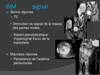
ONA : IRMsignes non spécifiques • Épanchement intra-articulaire • ± Œdème du spongieux du col • Corrélé à la douleur T2
ONA : IRM • Formes bilatérales > 50% • Lésion asymptomatique débutante controlatérale T1 T2
IRM vs SCINTIGRAPHIE • Sensibilité et spécificité de 98% • Précocité des signes vs scintigraphie ?? • Sujet débattu : 1 ou 2 semaines après le début des signes • Sensibilité très proche
ONA : ÉTENDUE • Stabilisation rare • Évolution fréquente destruction articulaire arthrosique • Facteur pronostique ? • % surface articulaire T2
CONSENSUS ARCO • Classification de Ficat + clinique + IRM • 7 stades • Dont étendue nécrose (IIIa,b,c et IVa,b,c) • 15%, 15-30%, >30% surface articulaire Subcommittee of Nomenclature of the International Association on Bone Circulation and Bone Necrosis
ONA ? : IRM • ONA débutante • Plage de bas signal • Sous-chondrale • Focale • Pas de liseré T1
F52 T1 T2 • +1 mois
M49 • +1 mois T1 T1
DIAGNOSTICS DIFFÉRENTIELS • Algodystrophie • Fracture de contrainte • Arthrose débutante
T1 STIR T1GD
T1 T2
T1 T2
ALGODYSTROPHIE : IMPACTION ÉPIPHYSAIRE • Fine image linéaire sous-chondrale • Bas signal en T1 et T2 • Injection de gadolinium détection • Proches de la surface épiphysaire • Sans rejoindre l’os sous-chondral
ALGODYSTROPHIE : DÉFORMATIONS FOCALES DE LA PLAQUE SOUS-CHONDRALE • Discrètes • 30 % • Enfoncement spontané : segments antérosup ou supéroexterne • Fractures-compression T2
ALGODYSTROPHIE : DOUTE DIAGNOSTIQUE PERSISTANT ENTRE ALGODYSTROPHIE ET ONA • CONTRÔLE IRM À 1 MOIS • MISE EN DÉCHARGE
FRACTURE DE FATIGUE • FRACTURE PAR INSUFFISANCE OSSEUSE • LOCALISATIONS HABITUELLES • Col du fémur • Cotyle • Branche ilio ou ischio-pubienne • Aileron sacré
T1 T2
COXARTHROSE T2 T1
CONCLUSION • Diagnostic précoce : IRM, scintigraphie • IRM précision anatomique et spécificité • Rx standard : complication et bilan pré-opératoire