Download
1 / 20
200 likes | 427 Views
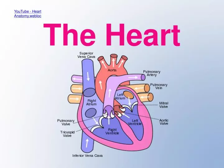
YouTube - Heart Anatomy.webloc. The Heart. Label your heart diagram with septum, L & R atria, L & R ventricles, two atrioventricular valves, pulmonary valve and aorta valve. Add blue and red arrows to show flow of oxygenated and deoxygenated blood. Heart Check.

E N D
YouTube - Heart Anatomy.webloc The Heart
Label your heart diagram with septum, L & R atria, L & R ventricles, two atrioventricular valves, pulmonary valve and aorta valve. • Add blue and red arrows to show flow of oxygenated and deoxygenated blood.
Heart Check • Why do the ventricles have thick walls? • Why do the atria have thin walls? • Why is the left ventricle thicker than the right? • How do the valves prevent backflow when the ventricle contracts?
Circulation and blood vessels • Veins towards heart • Arteries away • Label your diagram with the vena cava, pulmonary artery, pulmonary vein and aorta.
Circulation check • Which 2 vessels carry blood to the heart? • Which vessel carries deoxygenated blood to the lungs? • Which vessel carries oxygenated blood back to the heart? • Which vessel carries blood to the rest of the body? • “ Arteries carry oxygenated blood and veins carry deoxygenated blood ” True or False? Explain.
The Cardiac Cycle 1 2 3 3 phases: • Atrial systole (contracts) • Ventricular systole (contracts) • Diastole (relaxes) Concept II Practice- Rep#129DBC
Pressure changes in heart 1. What are phases a, b and c called? 2. Describe what is happening in the heart at points 1 – 10 on the graph. (c)
Cardiac Cycle check • What are the 3 phases of the cardiac cycle called? • Match them with phases 1,2 and 3 above. • Match ABC with phases 1,2 and 3 of cycle. Concept II Practice- Rep#129DBC
Coordinating the heartbeat • myogenic • SAN (sinoatrial node) the ’pacemaker’ • AVN (atrioventricular node) after slight delay • Bundle of His contraction from base of heart
Coronary Heart Disease • Coronary arteries provide heart muscle with blood carrying oxygen and glucose for respiration. • If these arteries become blocked (atheroma) problems arise. e.g. myocardial infarction
Atheroma • Fatty deposits build up under the endothelium of the artery when it becomes damaged. (damage can be caused by uneven blood flow, high blood pressure, chemicals or viral infection.) • White blood cells collect under the endothelium and absorb fatty materials e.g. LDLs ( contain cholesterol).
Atheroma • Lumen reduced.
Thrombosis • As a result of atheroma a lumpy area, called a plaque, forms on the artery wall. • This can lead to a blood clot forming (thrombosis). Can completely block the lumen.
Aneurysm • The artery wall can bulge in weakened areas. • This is an aneurysm. • It can burst. • Aneurysms and thrombosis can form in other parts of body too.
Atheroma can reduce blood supply to the heart muscle. • Angina is chest pain caused by exercise.
Myocardial Infarction • Muscle can die (infarction).
Symptoms of Myocardial Infarction • Severe pain in chest • Sick, breathless • Rapid but weak pulse • The affected area of muscle will stop working and the heartbeat can be disrupted which leads to uncoordinated contractions : fibrillation. • No pulse detected as not enough force to pump blood into aorta. • Emergency defibrillation is necessary. • 1/3 victims die within an hour : need heart compressions and artificial respiration until defibrillater available.
CHD worldwide • What are the risk factors?
Risk Factors • Age and Sex • Genetic factors • Smoking • High blood pressure • High concentration of LDLs (diet high in saturated fats)