Download
1 / 28
280 likes | 377 Views
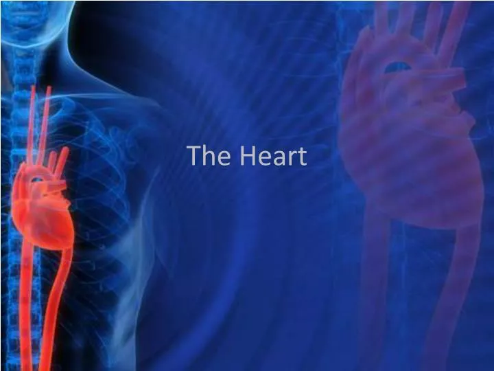
The Heart. Cardiovascular System. Consists of the heart and blood vessels Circulates blood in order to transport oxygen, nutrients, and wastes. The Organ. Located beneath 2 nd rib Avg. size: 14cm long 9 cm wide Base – attached to blood vessels Apex – points down & left. The Organ.

E N D
Cardiovascular System • Consists of the heart and blood vessels • Circulates blood in order to transport oxygen, nutrients, and wastes
The Organ • Located beneath 2nd rib • Avg. size: 14cm long 9 cm wide • Base – attached to blood vessels • Apex – points down & left
The Organ • Has a “skeleton” • Rings of dense connective tissue • Covered by a sac = pericardium • Consists of dense connective tissue surrounding serous membrane • Space in-between = pericardial cavity • Contains fluid to reduce friction
The Walls Consists of 3 walls: • Epicardium = connective tissue & epithelium • Blood & lymph capillaries, nerve fibers • Myocardium = cardiac muscle • contraction • Endocardium = epithelium & connective tissue • Blood vessels, specialized muscle fibers • Continuous with inner lining of vessels
The Chambers • 4 chambers • Top 2 = atria • Thin walls, receive blood • Separated by interatrial septum • Bottom 2 = ventricles • Thicker walls, force blood out • Separated by interventricular septum
The process – Part 1 • Right atrium receive blood from tissues • From superior & inferior vena cava • Low O2 • Right atrium contracts • Tricuspid valve allows blood from right atrium into the right ventricle • 3 flaps • Prevents backflow as well
The process – Part 1 • Right ventricle contracts • Tricuspid valve closes • Pulmonary valve opens • Exit called the pulmonary trunk • Trunk splits into right and left arteries and takes blood to lungs
The process – Part 2 • Left atrium receive blood from lungs • 4 pulmonary veins • Left atrium contracts • Mitral valve allows blood from left atrium into the left ventricle • 2 flaps • Prevents backflow as well
The process – Part 2 • Left ventricle contracts • Mitral valve closes • Aortic valve opens • Exit into aorta • Left ventricle relaxes which closes aortic valve • Aorta branches to distribute blood throughout the body
Valve Control • Fibrous strings called chordaetendineae prevent the flaps of the mitral and tricuspid valves from swinging backwards into the atria