Download
1 / 26
680 likes | 1.52k Views
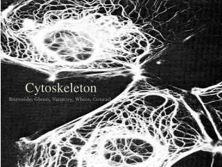
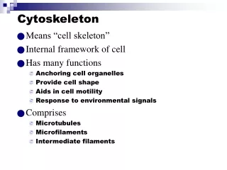
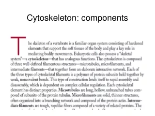
Lecture 14 Cytoskeleton: components. Cytoskeleton proteins revealed by Commassie staining. Cytoskeleton: filament system. Three types of filaments and accessory proteins (assembly of cytoskeleton, motor proteins that move organelles or filaments). Internal order Shape and remodel surface

E N D
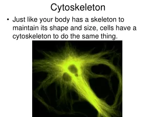
Lecture 14 Cytoskeleton: components
Cytoskeleton proteins revealed by Commassie staining Cytoskeleton: filament system Three types of filaments and accessory proteins (assembly of cytoskeleton, motor proteins that move organelles or filaments) Internal order Shape and remodel surface Move organelles Movement Cell division
Dynamic and adaptable Actin filaments: shape of the cell’s surface and whole cell locomotion 5-9 nm diameter Microtubules: positions of membrane- enclosed organelles, intracellular transport 25 nm diameter Intermediate filaments: mechanical strength and resistance to shear stress 10 nm diameter
Cytoskeletal filaments are all constructed from smaller protein subunits Intermediate filaments: smaller elongated and fibrous subunits Actin and microtubule filaments: compact and globular subunits All form as helical assemblies of subunits Noncovalent interactions: rapid assembly and disassembly
Multiple protofilaments give strength and adaptability Ends are dynamic
Intermediate filaments are resistent to bending or stretching forces
The structure of a microtubule and its subunits hollow and cylindrica and polar GTP! GTP heterodimer 13 parallel protofilaments
The structure of an actin monomer and actin filament two parallel protofilaments that twist around each other in a right-handed helix polar ATP monomer Flexible but cross-linked and bundled together by accessory proteins in a living cell
The preferential growth of microtubules at the plus end Plus end: polymerize and depolymerize faster than minus end Microtubules: Plus end- b subunit Minus end- a subunit Actin filaments Plus end- barbed end Minus end- pointed end
The treadmilling of an actin filament Structural difference between the two ends D form polymer leans towards disassembly
Treadmilling behavior of a microtubule as in a living cells The extent of treadmilling inside the cell Is uncertain. Actin treadmilling is observed in vitro. A treadmilling-like phenomenon is seen in living cells for microtubules Tubulin conjugated with fluorescent dye 1/20 subunit is fluorescent “Microtubule lattice”
Dynamic instability:predominant in microtubules GTP hydrolysis “catch up” Treadmilling: predominant in actin filaments
GTP hydrolysis causes filament to curve
Lateral bonds force GDP-containing protofilaments into a linear conformation
Direct observation of the dynamic instability of microtubules in a living cell Dynamic instability of individual actin filaments cannot be observed readily-difference between two ends are not so extreme However the actin filament turn over is rapid: individual filament persists for a only few minutes
The dynamic behavior of filaments allows cells to change structures rapidly and Giant multinuclear cell of a fly early embryo Actin filaments:red Mircotubules:green One division per 10 minutes
Actin and tubulin are highly conserved: they have to bind to many proteins directly and indirectly Accessory proteins and intermediate filament proteins are not as conserved
A model of intermediate filament construction Intermediate filaments are only found in some metazoans:vertebrates, nematodes,molluscs Parallel Antiparrel Not required in every cell type Ancesters: nuclear lamins “subunit” No polarity! Easily bent Hard to break 8 parallel protofilaments
Mechanical properties of actin, tubulin and Intermediate filament polymers viscometer Microtubules: easily deformed and then rupture Actin filaments are more rigid and also rupture easily Intermediate filaments: easily deformed and don’t rupture--maintain cell integrety
Intermediate filaments impart mechanical stability to animal cells Keratin filaments in epithelial cells The most diverse family 20 in human epithelial cells 10 more in hair and nails Diagnosis of epithelial cancers (carcinomas) “desmosomes”
Blistering of the skin caused by mutant keratin genes Epidermolysis bullosa simplex:the skin blisters in response to very slight mechanical stress Truncated keratin (missing both the N- C- domains) Tg mice Other blistering diseases: mouth, esophageal lining and cornea of the eye-- mutations of different keratins
Two types of intermdiate filaments in cells of the nervous system Neurofilaments:axons Regular spacing NF-L, NF-M, NF-H proteins coassemble glia axon NF-M and NF-H have long C-terminal tails That bind to neighboring filaments:uniform spacing When axons grow, subunits are added at the filament ends and along the filament length; axon diameter increase 5 fold In ALS (Lou Gehrig’s Disease), there is an accumulation and abnormal assembly of Neurofilaments in motor neuron cell bodies and axon--interfere with normal axon transport
Actin filaments and microtubules are targets of many plant toxins Amanita Phalloids (death cap) (Eat raw meat)
Effect of the drug taxol on microtubule organization treatment of cancers
Summary Three types of cytoskeletal filaments, protofilaments; Subunits, polymerization, treadmilling, dynamic instability; Intermediate filaments, cell integrity, diseases caused by mutations in the intermediate filament genes 4. Natural toxins and cytoskeleton