Download
1 / 17
210 likes | 523 Views
Cytoskeleton. Yasir Waheed. Cells have to organize themselves in space and interact mechanically with their environment. They have to be correctly shaped, physically robust, and properly structured internally.

E N D
Cytoskeleton YasirWaheed
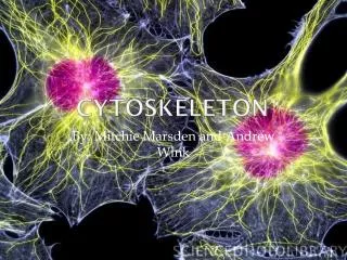
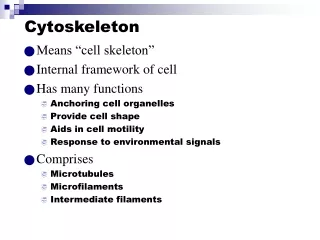
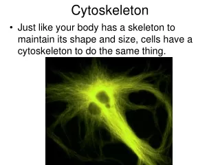
Cells have to organize themselves in space and interact mechanically with their environment. • They have to be correctly shaped, physically robust, and properly structured internally. • Many of them also have to be able to change their shape and move from place to place. All of them have to be able to rearrange their internal components as they grow, divide, and adapt to changing circumstances. • All these spatial and mechanical functions are developed to a very high degree in eucaryotic cells, where they depend on a remarkable system of filaments called the cytoskeleton Figure 16-1. The cytoskeleton. A cell in culture has been fixed and stained with Coomassie blue, a general stain for proteins. Note the variety of filamentous structures that extend throughout the cell.
The cytoskeleton pulls the chromosomes apart at mitosis and then splits the dividing cell into two. • It drives and guides the intracellular traffic of organelles, ferrying materials from one part of the cell to another. • It supports the fragile plasma membrane and provides the mechanical linkages that let the cell bear stresses and strains without being ripped apart as the environment shifts and changes. • It enables some cells, such as sperm, to swim, and others, such as fibroblasts and white blood cells, to crawl across surfaces. • It provides the machinery in the muscle cell for contraction and in the neuron to extend an axon and dendrites. • It guides the growth of the plant cell wall and controls the amazing diversity of eucaryotic cell shapes. • The various functions of the cytoskeleton is performed by three families of protein molecules , which assembles to from three type of filaments.
The Self-Assembly and Dynamic Structure of Cytoskeletal Filaments Three types of cytoskeleton filaments are common to many eukaryotic cells. Intermediate filaments provide mechanical strength and resistance to shear stress. Microtubulesdetermine the positions of membrane-enclosed organelles and direct intracellular transport. Actin filaments determine the shape of the cell's surface and are necessary for whole-cell locomotion. But these cytoskeletal filaments would be ineffective on their own. Their usefulness to the cell depends on a large number of accessory proteins that link the filaments to other cell components, as well as to each other. The set of accessory proteins is essential for the controlled assembly of the cytoskeletal filaments in particular locations, and it includes the motor proteins that either move organelles along the filaments or move the filaments themselves.
Actin filaments • Actin filaments (also known as microfilaments) are two stranded helical polymers of the protein actin. • They appear as flexible structures, with a diameter of 5-9nm. • They are organized into a variety of linear bundles. • Actin filaments are dispersed throughout the cell, they are mostly concentrated in the cell cortex, just beneath the plasma membrane.
Microtubles • Microtubles are the long, hollow cylinders made of the protein tubulin • They have diameter of 25nm and are much more rigid than actin filaments. • Microtubles are long and straight and typically have one end attached to a single microtuble organizing center (MTOC) called centrosome.
Intermediate Filaments • Intermediate Filaments are the rope like fibers with a diameter of around 10nm; they are made up of intermediate filament proteins , which constitute a large and heterogenous family. • One type of the intermediate filament forms a mesh work called the nuclear lamina just beneath the inner nuclear membrane. • Other types extend across the cytoplasm, giving cell mechanical strength. • In an epithelial tissue, they span the cytoplasm from one cell-cell junction to another, thereby strengthening the entire epithelium.
Figure 16-2. The cytoskeleton and changes in cell shape. The formation of protein filaments from much smaller protein subunits allows regulated filament assembly and disassembly to reshape the cytoskeleton. (A) Filament formation from a small protein. (B) Rapid reorganization of the cytoskeleton in a cell in response to an external signal.
Filaments Formed from Multiple Protofilaments Have Advantageous Properties • The linking of protein subunits together to form a filament can be thought of as a simple association reaction. A free subunit binds to the end of a filament that contains n subunits to generate a filament of length n + 1. • The addition of each subunit to the end of the polymer creates a new end to which yet another subunit can bind. • However, the robust cytoskeletal filaments in living cells are not built by simply stringing subunits together in this way in a single straight file. • A thousand tubulin monomers, for example, lined up end to end, would be enough to span the diameter of a small eucaryotic cell, but a filament formed in this way would not have enough strength to avoid breakage by ambient thermal energy, unless each subunit were bound very tightly to its neighbor. Such tight binding would limit the rate at which the filaments could disassemble, making the cytoskeleton a static and less useful structure.
Figure 16-3. The thermal stability of cytoskeletal filaments with dynamic ends. Formation of a cytoskeletal filament from more than one protofilament allows the ends to be dynamic, while the filaments themselves are resistant to thermal breakage. In this hypothetical example, the stable filament is formed from five protofilaments. The bonds holding the subunits together in the filaments are shown in red.
Figure 16-6. The structure of a microtubule and its subunit. (A) The subunit of each protofilament is a tubulin heterodimer, formed from a very tightly linked pair of alpha and beta tubulin monomers. The GTP molecule in the alpha-tubulin monomer is so tightly bound that it can be considered an integral part of the protein. The GTP molecule in the beta-tubulin monomer, however, is less tightly bound and has an important role in filament dynamics. Both nucleotides are shown in red. (B) One tubulin subunit (alpha-beta heterodimer) and one protofilament are shown schematically. Each protofilament consists of many adjacent subunits with the same orientation. (C) The microtubule is a stiff hollow tube formed from 13 protofilaments aligned in parallel. (D) A short segment of a microtubule viewed in an electron microscope. (E) Electron micrograph of a cross section of a microtubule
Figure 16-11. Dynamic instability due to the structural differences between a growing and a shrinking microtubule end • A single microtubule end may undergo transitions between a growing state and a shrinking state. A growing microtubule has GTP containing subunits at its end, forming a GTP cap. If nucleotide hydrolysis proceeds more rapidly than subunit addition, the cap is lost and the microtubule begins to shrink, an event called a "catastrophe." But GTP-containing subunits may still add to the shrinking end, and if enough add to form a new cap, then microtubule growth resumes, an event called "rescue.“ • (B) Model for the structural consequences of GTP hydrolysis in the microtubule. The addition of GTP containing tubulin subunits to the end of a protofilament causes the end to grow in a linear conformation that can readily pack into the cylindrical wall of the microtubule. Hydrolysis of GTP after assembly changes the conformation of the subunits and tends to force the protofilament into a curved shape that is less able to pack into the microtubule wall. • (C) In an intact microtubule, protofilaments made from GDP-containing subunits are forced into a linear conformation by the many lateral bonds within the microtubule wall, given a stable cap of GTP-containing subunits. Loss of the GTP cap, however, allows the GDP-containing protofilaments to relax into their more curved conformation. This leads to a progressive disruption of the microtubule. Above the drawings of a growing and a shrinking microtubule, electron micrographs show actual microtubules in each of these two states. Note particularly the curling, disintegrating GDP-containing protofilaments at the end of the shrinking microtubule.
Figure 16-16. A model of intermediate filament construction. The monomer shown in (A) pairs with an identical monomer to form a dimer (B), in which the conserved central rod domains are aligned in parallel and wound together into a coiled coil. (C) Two dimers then line up side by side to form an antiparallel tetramer of four polypeptide chains. The tetramer is the soluble subunit of intermediate filaments. (D) Within each tetramer, the two dimers are offset with respect to one another, thereby allowing it to associate with another tetramer. (E) In the final 10-nm rope-like filament, tetramers are packed together in a helical array, which has 16 dimers in cross-section. Half of these dimers are pointing in each direction. An electron micrograph of intermediate filaments are shown on the upper left.
Figure 16-23. The centrosome. (A) The centrosome is the major MTOC of animal cells. Located in the cytoplasm next to the nucleus, it consists of an amorphous matrix of protein containing the gamma-tubulin ring complexes that nucleate microtubule growth. This matrix is organized by a pair of centrioles, as described in the text. (B) A centrosome with attached microtubules. The minus end of each microtubule is embedded in the centrosome, having grown from a gamma-tubulin ring complex, whereas the plus end of each microtubule is free in the cytoplasm.
Figure 16-26. A microtubule array can find the center of a cell. After the arm of a fish pigment cell is cut off with a needle, the microtubules in the detached cell fragment reorganize so that their minus ends end up near the center of the fragment, buried in a new microtubule-organizing center.