Download
1 / 4
40 likes | 170 Views
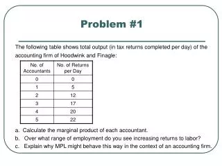
Problem 1. Among 4.5 million births in one population during a period of 40 years, 41 children diagnosed with the autosomal dominant condition aniridia were born to normal parents

E N D
Problem 1 • Among 4.5 million births in one population during a period of 40 years, 41 children diagnosed with the autosomal dominant condition aniridia were born to normal parents • a. Assuming that these cases were due to new mutations, what is the estimated mutation rate at the aniridia locus? On what assumptions is this estimate based, and why might this estimate be either too high or too low? Note: problem 1 from chapter 6 page 94
Answer Problem 1 • a. Assuming that these case were due to new mutations, what is the estimated mutation rate at the aniridia locus? On what assumptions is this estimate based, and why might this estimate be either too high or too low? • Assume 40 years is one generation. Then 41 mutations/9 million alleles=4.55x10⁻⁶. This is based on the following assumptions • ascertained cases result from new mutations • the disease is fully penetrant • all new mutant are liveborn and ascertained • only a single locus at which the mutation can lead to anirida • If there are multiple loci • then the estimate is too high • If some mutation are not ascertained (because of death or lack of penetrance) • the estimate is to low Note: problem 1 from chapter 6 page 94
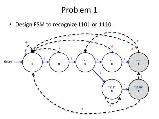
Problem 2 • The gel to the left is a diagnostic method used to determine the size of the CGG repeat in the FMR1 gene. The positions of normal alleles are indicated by the white arrows while the positions of diagnostic alleles are indicated by the gray arrows • Lane 1 is a Normal male 2.8 Kbp unmethylated EcoR1/Eag1 fragment. • Lane 2 is a Premutation male Slightly increased size 2.8 Kbp EcoR1/Eag1 fragment due to slight increase in CGG repeat number. • If lanes 3 and 4 are males do they have fragile X syndrome. If they are affected which males do you thinks has the most sever symtoms. • Lane 5 is a Normal female 5.2 Kbp methylated (inactive X chromosome) and 2.8 Kbp unmethylated (active X chromosome) EcoR1/Eag1 fragments. • Lane 6: Premutation carrier female Slightly increased size 2.8 Kbp and 5.2 Kbp EcoR1/Eag1 fragments due to slight increase in the FMR1 CGG repeat number carried by one X chromosome. Some methylated and some unmethylated. • If Lanes 7 and 8 are females do they have fragile X syndrome. If they are affect who do you think would have the most sever degree of impairment..
Answer Problem 2 In gel electrophoresis larger DNA fragments travel slower in the gel. Therefore small DNA fragments will be closer to the bottom of the gel and larger DNA fragments will be closer to the top. • The gel to the left is a diagnostic method used to determine the size of the CGG repeat in the FMR1 gene. The positions of normal alleles are indicated by the white arrows while the positions of diagnostic alleles are indicated by the gray arrows • Lane 1 is a Normal male This indicates the size of a FMR1 fragment for an unaffected male • Lane 2 is a Premutation male This indicates the size of a FMR1 fragment for an unaffected male whose children are likely to receive a fragile X chromosome when the repeat expands • If lanes 3 and 4 are males do they have fragile X syndrome. • The individual in lanes 3 and 4 have very large fragment. They almost certainly have fragile X syndrome. • If they are affected which males do you thinks has the most sever symtoms. • The individual in has the largest fragment so may be the most severely affected. • Lane 5 is a Normal female 5.2 Kbp methylated (inactive X chromosome) and 2.8 Kbp unmethylated (active X chromosome) These are the size of the fragments for an unaffected female • Lane 6: Premutation carrier female Slightly increased size 2.8 Kbp and 5.2 Kbp EcoR1/Eag1 fragments due to slight increase in the FMR1 CGG repeat number carried by one X chromosome. Some methylated and some unmethylated. These are the fragment size for a female who is unsymptomatic but whose children will have a larger repeat size and are more likely to be affected • If Lanes 7 and 8 are females do they have fragile X syndrome. • Both have much larger repeat sizes and are at least carrier of the disease. In all likely hood they are mildly affect • If they are affect who do you think would have the most sever degree of impairment.. • Difficult to tell • The female in lane 8 has the largest repeat but one normal chromosome. This person may have the most sever symptoms • The female in lane 7 has two affected chromsomes but the repeat sizes are small. Because both chromosomes are affected she may have more sever symptoms than individula 7. In this gel we are looking at DNA fragments from the 5’ region of the FMR1 gene. The larger the DNA fragment the larger the CGG repeat. In fact the difference in the size of the DNA fragments is caused by changes in the CGG repeat size.