Download
1 / 14
140 likes | 229 Views
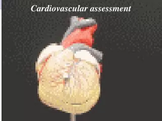
Assessment of the Cardiovascular System. p.343-348. Ways to Assess the Heart’s Condition. Measuring Pulse and Blood Pressure Listening to Heart Sounds Determining Cardiac Output Measuring Muscle activity with electrocardiography Inserting a cardiac catheter Using echocardiography. Pulse.

E N D
Assessment of the Cardiovascular System p.343-348
Ways to Assess the Heart’s Condition • Measuring Pulse and Blood Pressure • Listening to Heart Sounds • Determining Cardiac Output • Measuring Muscle activity with electrocardiography • Inserting a cardiac catheter • Using echocardiography
Pulse • Defined: blood surge against wall • Found in arteries close to the skin • Most common is radial • Healthy range = 60-80 bpm
Blood Pressure • Systolic = contraction of the ventricles • Diastolic = ventricle relaxation • Normal BP= 120/80 (systolic/diastolic) • Healthy systolic is less than 140 and greater than 90 • Healthy diastolic should be less than 100
Heart Sounds • Lubb Sound • Heard first • Mitral and tricuspid valves closing between the atria and ventricles • Dupp Sound • Heard second • Shorter and higher pitched • Closing of the aortic and pulmonary valves as blood is pumped out of the heart • Murmurs • Abnormal or extra sounds http://depts.washington.edu/physdx/heart/demo.html
Cardiac Output • Heart controls heart rate • Heart rate = rate at which blood circulates to the tissues • Blood that is in ventricles is pumped through arteries with each contraction • Cardiac output= stroke volume ml/beat X HR beats/min. • Normal cardiac output = 4-8 L / min. • Can affect the BP • Too high or too low = indication of a cardiovascular system disorder
Picture of non-invasive cardiac output measurement technique.
Electrocardiogram (EKG) • Defined: record of the electrical activity of the myocardium • Electrodes attach to body and measure electrical changes • Each part of the EKG pattern indicates specific electrical activity
Echocardiography (ECG) • Defined: an ultrasonic procedure used to evaluate the structures and motion of the heart • Using ultrasonic waves • Echoes • Used to detect mitral valve defects and atrial tumors
Cardiac Catheterization p. 804 • Defined: placement of catheter through a vein or artery and into the heart • Dye is released and traced using an x-ray • Used for: • Measuring pressure in the chambers • Taking blood samples • View obstruction in the vessels