Download
1 / 67
670 likes | 810 Views
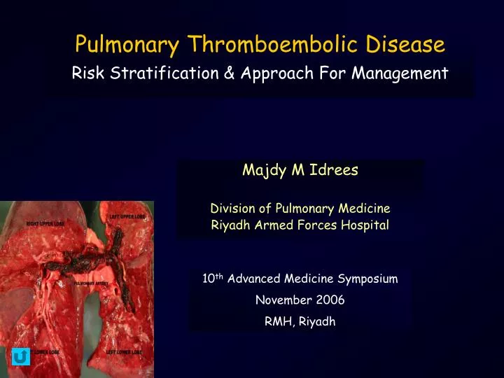
Pulmonary Thromboembolic Disease Risk Stratification & Approach For Management. Majdy M Idrees Division of Pulmonary Medicine Riyadh Armed Forces Hospital. 10 th Advanced Medicine Symposium November 2006 RMH, Riyadh. An Expert:. A man who has stopped thinking:. He Knows!!.

E N D
Pulmonary Thromboembolic DiseaseRisk Stratification & Approach For Management Majdy M Idrees Division of Pulmonary Medicine Riyadh Armed Forces Hospital 10th Advanced Medicine Symposium November 2006 RMH, Riyadh
An Expert: A man who has stopped thinking: He Knows!!
Pulmonary Embolism Risk Stratification & Approach of Management “Venous thrombosis is always a severe disease and is often fatal, because fragments of the thrombi may detach and occlude branches of the pulmonary artery... the occlusion of the main branches of the pulmonary artery causes a striking rise of the blood pressure in these vessels. This rise, which the right heart might fight in order to ensure circulation, may sometimes lead to cardiac arrest.” Picot 1884 Lecons de Clinique Médicale
Pulmonary Embolism Risk Stratification & Approach of Management Definition It is a clinical syndrome of high mortality characterized by acute pulmonary arterial occlusion with resultant sudden elevation in pulmonary artery pressure and right ventricular failure
Pulmonary Embolism Risk Stratification Pulmonary Embolism Risk Stratification Simple Complicated Submassive Massive
Complicated Pulmonary Embolism Massive Pulmonary Embolism Visual Scoring Functional Scoring • Perfusion defect >50% • Filling defect / obstruction in 2 lobar arteries • Obstruction of main (R/L) PA 1. Severe refractory hypoxic 2. Hemodynamic instability 3. Acute RV failure 4. Tissue hypoperfusion (low urine output)
Predicting PE Perfusion Defects < 30% 30% - 50% >50% Galle Thromb Haemost 2001; 86:1156-60
PE Severity: Echo Assessment % Obstruction Mastora Eur Radiol 2003; 13:29-35
CT Severity Assessment Non-severe PE Severe PE (Heparin tx) (Lysis/Embolectomy) Collomb Eur Radiol 2003; 13:1508-14
Pathophysiology Obstruction RV Pressure Load Neuro-hormonal Decreased RV CPP Decrease RV Output RV Decompensation Ischemia Increase RV Volume • VO2 • Wall stress • Septal Shift • Pericardial Restriction COP / MAP Decrease LV Distensiblity Decreased LV Preload
Vascular Obstruction and Acute Pulmonary Hypertension 70 50 Pulmonary Vascular Obstruction Angiogram (%) 30 10 10 30 20 40 Pulmonary Arterial Mean Pressure (mmHg) McIntyre Am J Card 1971; 28:288-294
30 20 10 0 10 20 30 Vascular Resistance vs Obstruction TPVR (mmhg.1.min.m) • • • • • • • • • • • • • ° ° • • • • • • ° ° • • • ° • ° ° ° Miller index Petitpretz Circ 1984; 70:861-866.
Intervention Diagnostic – Therapeutic Approach
Outcome in Pulmonary Embolism Risk Stratification 100 70 30 10 0 Sudden Death Cardiac Arrest Infliction Point Mortality Shock Hemodynamically Stable & RV Normal Severity Embolism size Cardiopulmonary Status
Outcome in Pulmonary Embolism Risk Stratification 100 70 30 10 0 Sudden Death Cardiac Arrest Mortality Shock Hemodynamically Stable & RV Normal Severity Embolism size Cardiopulmonary Status
Sudden Death in Massive PE • Mortality is almost 100% • No intervention measure has proven effective
Outcome in Pulmonary Embolism Risk Stratification 100 70 30 10 0 Sudden Death Cardiac Arrest Mortality Shock Hemodynamically Stable & RV Normal Severity Embolism size Cardiopulmonary Status
Echo and Cardiac Arrest • 48 patients in/out hospital cardiac arrest • Diagnosis obtained via TEE • Myocardial infarction 21 • Cardiac tamponade 6 • Pulmonary embolism 6 • Aortic dissection/rupture 5 • Papillary muscle rupture 1 • Absence of cardiac structural abnormalities 7 • Other diagnosis 2 • Sensitivity 93% specificity 50%, positive predictive value 87% • 31% major therapeutic decisions based upon TEE JACC 30:780-783, 1997
Thrombolysis in CPR • Majority of PE deaths within 1st hour of symptoms • Bolus thrombolysis in arrest- “Reported” • Initiated after failure of conventional CPR • Stabilization • Minimal bleeding complications • Microcirculatory Reperfusion 77% Stabilized 67% Survived Bottiger Fibrinolysis and Proteolysis, 1997
Outcome in Pulmonary Embolism Risk Stratification 100 70 30 10 0 Sudden Death Cardiac Arrest Mortality Shock Hemodynamically Stable & RV Normal Severity Embolism size Cardiopulmonary Status
The Aphorisms of Hippocrates “In acute diseases,,, coldness of the extremities,, is a very bad sign.”
UPET 1970 Heparin Thrombolysis 36% 6% Alpert 1976 Heparin Venous Ligation 25% 5% Miller 1977 Heparin Thrombolysis Embolectomy 22% 11% Verstraete 1988 Thrombolysis 14% 4% Tilsner 1991 Thrombolysis 40% 2% Pulmonary EmbolismShock vs. Non-Shock Mortality Shock Mortality Non-Shock Mortality Study Treatment
Major Pulmonary Embolism Diagnostic & Therapeutic Approach Shock • Emboli in PA • AMI / Aortic Dissection / Tamponade Start Heparin ECHO TEE/TTE Certain Diagnoses • Spiral CT • V/Q • Angio Yes Establish Diagnosis RV Pressure Overload? No - + No • Alternative DX • Resuscitate & Stabilize Embolectomy Peruse other dx Lysis Candidate Lytic Rx Yes
Measures to Improve Hemodynamics • Flow (Cardiac Output)
Heparin Therapy Exclusion Criteria PIOPED “Patients with shock or major disability due to Pulmonary Embolism were excluded because random assignment to a placebo group was considered unethical.” PIOPED Investigators CHEST 1990; 97:528-33
Management Massive PE With Shock Heparin Therapy • The efficacy of heparin is attributed to an impairment of clot propagation and the prevention of recurrent PE • Recurrent PE is reported to be the most common cause of death in hemodynamically stable patients • Prog Cardiovasc Dis17,257-270 • An inability to establish an early therapeutic level for aPTT is associated with a higher rate of recurrence and impairs the efficacy of anticoagulation therapy with warfarin • It is recommended that heparin therapy be given for 7 to 10 days and that the initiation of warfarin therapy be delayed until the aPTT is at a therapeutic level for 3 days.
Thrombolists Perspective – “Works Great” • Hastens thromboembolic resolution • Removes pulmonary thromboemboli more completely • Hastens dissolution of thrombi in legs • May decrease mortality from pulmonary embolism • May diminish the incidence of chronic thromboembolic pulmonary hypertension Sasahara J Cardiovascular Medicine 1980
Traditionalists Perspective – “Unfulfilling” • Does fibrinolytic therapy decrease mortality in acute PE? • What is the impact of fibrinolytic therapy on short and long term recovery from acute PE? • Does fibrinolytic therapy decrease the rate of recurrent PE? • How do the complicationsand Cost of fibrinolytic therapy compare with those of heparin? • “Available data does not support the FDA conclusion that fibrinolytic agents are indicated for massive PE… there is insufficient data to determine whether the second approved indication for fibrinolytic therapy – PE accompanied by failure to maintain BP without supportive measures – is appropriate.” Dalen J Cardiovascular Medicine 1980
Accelerated Clot Lysis Recurrent Embolism Angiograms Perfusion scans Chronic Pul HTN Pul capillary blood volume Quality of Life Hemodynamic Improvement Pulmonary pressures Symptoms Echocardiogram Mortality Thrombolytic TherapyPutative Benefits PROVEN SPECULATIVE
Lytic Heparin UPET ‘73 UK 24 24.1%* 8.3% % Scan defect Tibbutt ‘74 SK 72 -13.3* -2.8 Δ Angio severity Ly ‘78 SK 72 -11.3* -3.4 Δ Angio severity PIOPED ‘90 rt-PA 24 10% 0% % Δ mismatch scan defect Levine ‘90 rt-PA 24 34.4% 12.0% % showing 50% improvement scan PAIMS 2 ‘92 rt-PA 2 -3.5* -0.1 Δ Angio severity Goldhaber ‘93 rt-PA 24 14.6%* 1.5% % scan Study/Yr Agent Time Post Resolution Metric Early Resolution Rate Lytic vs Heparin
Angiographic Severity UPET Complete (91-100%) 4 UPET JAMA 1970; 214:2163-2172 Marked (61-90%) 3 Severe 3 N = 57 N = 57 Moderate (31-60%) 2 Moderate 2 Slight Improvement ( 30%) 1 Minimal 1 No change 0 Normal 0 Heparin Urokinase Heparin Urokinase 24 Hours Baseline
Rate and Extent Clinical-Hemodynamic Resolution Reported in UPET Urokinase Heparin 24 hours Δ Baseline Baseline 24 hours Δ UPET JAMA 1970; 214:2163-2172
Total Pulmonary Resistance Evolution Heparin 0 -20 Streptokinase % change from baseline -40 rt-PA -60 10 8 0 4 6 12 2 Time (h) Meneveau European Heart Journal 1997; 18:1141-1148
Right Ventricular Ejection Fraction Evolution 80 70 60 rt-PA 50 40 % change from baseline Streptokinase 30 20 10 Heparin 10 8 0 4 6 12 2 Time (h) Meneveau European Heart Journal 1997; 18:1141-1148
Recurrent PE With Thrombolytic Therapy Heparin Lytic Study Agent Dalen Venous Thromboembolism Lung Biology in Health and Disease 2003
Thrombolytic Therapy Hemorrhagic Complications • Major hemorrhage 8-12% • Similar amongst agents • Fatal hemorrhage 1-2% • Intra-cranial hemorrhage 1.2-2.1% • Fatal in 50% Arcasoy Chest 1999; 115:1695-1707
Management Massive PE With Shock • Surgical Embolectomy • Allows rapid and complete removal of the clot • Survival rate in retrospective review 40-60% • Ann Thorac Surg 1991 • Cardiac arrest is the most preoperative prognostic factor • Indicated when • Thrombolysis is contraindicated or failed • “Unyeilding” hypotension despite max therapy for > 1 h • Ongoing/intermittent cardiac arrest
Infliction PointEmboli in TransitRV Dysfunction Submassive Pulmonary Embolism
Outcome in Pulmonary Embolism Risk Stratification 100 70 30 10 0 Sudden Death • Infliction Point • RV Dysfunction • Troponin T • NDH Cardiac Arrest Mortality Shock Hemodynamically Stable & RV Normal Severity Embolism size Cardiopulmonary Status
Isolated RV Dysfunction • RV dysfunction has long been recognized as a marker for poor outcome in patients with PE, especially in those with hemodynamic instability • RV dysfunction in hemodynamically stable patients has been identified as a predictor of worse outcome and appears to be related to the presence of recurrent PEs. • 10% of hemodynamically stable patients with RV dysfunction will deteriorate into shock with a 50% mortality rate attributed to those with recurrent PEs
Massive Pulmonary Embolism Prognostic Factors - ICOPER Hazard Ratio Factor Goldhaber Lancet 1997; 353: 1386-1389
Outcomes Hemodynamically Stable Confirmed PE with RV Dysfunction Tx Heparin Study Year n PE Deaths % Mortality
Alteplase vs Heparin Acute PE Assessing RV Function and Perfusion in Hemodynamically Stable Patients Rt-PA Heparin All RV Dysfxn 2 fatal 3 non-fatal Goldhaber Lancet 1993; 341:507-11
Major Pulmonary EmbolismThrombolytic Therapy - MAPPET RV Dysfxn + BP-No pressors (719) Lysis (24%) Heparin (76%) Mortality 4.7% 11.1% Recurrent PE 7.7% 18.7% Major bleed 21.9% 7.8% • Clinical factors death: syncope, BP, CHF, COPD • Primary lysis independent predictor of survival • Caution!! Heparin group old, CHF, COPD Konstantinides Circ 1997; 96:882-888
Non-shock Mortality Thrombolytic Therapy Mortality Patients Heparin Lytic % (N) Lytic Heparin % (N) Study