Download
1 / 87
870 likes | 905 Views
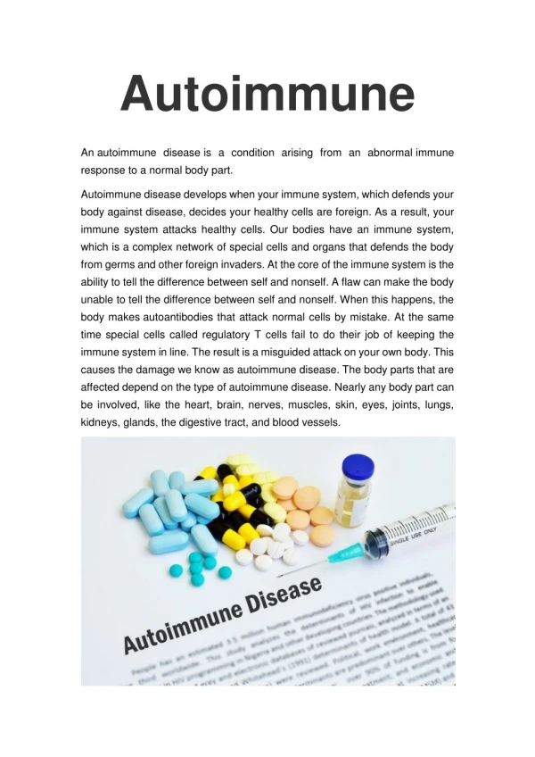
Autoimmune Hepatitis. -Chronic hepatitis with immunologic abnormalities -Histologic features are similar to chronic viral hepatitis -Indolent or severe course -Dramatic response to immunosuppressive therapy. Features: 1-Female predominance (70%) 2-Negative serelogy for viral Ags.

E N D
Autoimmune Hepatitis -Chronic hepatitis with immunologic abnormalities -Histologic features are similar to chronic viral hepatitis -Indolent or severe course -Dramatic response to immunosuppressive therapy
Features: 1-Female predominance (70%) 2-Negative serelogy for viral Ags. 3-↑serum Ig (>2.5 g/dl) 4-High titers of autoantibodies (80% of cases) 5-The presence of other autoimmune diseases as RA, thyroiditis, sjogern syndrome, UC in 60% of the cases
The type of autoantibodies 1-Antismooth muscle Abs anti actin anti troponin anti tropomyosin 2-liver/kidney microsomal Abs anti cytochrome P-450 components anti UDP-glucuronosyl transferases 3-Anti – soluble liver / pancreas antigen
Outcome Mild to severe chronic hepatitis Full remission is unusual Risk of cirrhosis is 5% which is the main cause of death
Nonalcoholic Fatty Liver Disease Types: 1.Steatosis ( Fatty liver) 2.Steatohepatitis hepatocyte destruction parenchymal inflammation progressive pericellular fibrosis
Predisposing factors : 1-Type 2 DM 2-Obesity : body mass index > 30 kg /m2 in caucasians > 25 kg /m2 in Asians 3-Dyslipidemia ( ↑ TG, ↑LDL, ↓HDL)
Pathogenesis .Metabolic syndrome . Insulin resistance . Obesity . Dyslipidemia
Mechanism of fatty accumulation 1.Impaired oxidation of fatty acids 2.Increased synthesis & uptake of FFA 3.Decreased hepatic sec. of VLDL . ↑TNF , IL6 , chemokine →liver inflammation & damage
Clinically -NAFLD is the most common cause of incidental ↑ in transaminases -Most pts. are asymptomatic -Non-specific symptoms Fatigue, malaise, RUQ discomfort -Severe symptoms -Liver Bx is required for dx. -NAFLD m.b a significant contributer to cryptogenic cirrhosis
Hemochsomatosis • Excessive accumalation of body iron (liver & pancreas) • 1ry or 2ry (genetic or acquired ) • Genetic hemochromatosis ( 4 variants) • The most common form is aut.recessive disease of adult onset caused by mutation in the HFE gene on chr.6
Causes of acquired hemosidrosis : 1-multiple transfusions 2-ineffective erythropoiesis (β-thalassemia ) 3-increased iron intake (Bantu sidrosis ) 4-chronic liver disease
Clinical Features: 1-Micronodular cirrhosis (all patients) 2-D.M ( 75 – 80%) 3-skin prigmentation 75-80%) 4-cardiomegaly (arrhythmias, cardiomyopathy) 5- joints disease 6- testicular atrophy
Symptoms appear 5th – 6th decades not before age 40 • M:F ratio 5 - 7: 1 • earlier clinical presentation in males partly because physiologic iron loss (menstruation, pregnancy) retards iron accumulation in women.
Pathogenesis -1ry defect in intestinal absorption of dietary iron. -Total body iron 2-6gm in adults 0.5gm in liver mostly in hepatocytes -In disease >50gm of iron accumulated → 1/3 in liver -There is a defect in regulation of intestinal absorption of dietary iron leading to net iron accumulation of 0.5 – 1 gm/yr.
HFE gene regulates the level of hepcidin hormone synthesized in liver • Hepicidin normally inhibits iron absorption. • When hepcidin levels are reduced there is increased iron absorption. • HFE gene deletion causes→ ↓Hepcidin levels→ iron overload
-Two mutations can occur in HFE gene: 1-Mutation at 845 nucleotide → tyrosine substitution for cystine at AA 282 ( C282 Y ) 2-aspartate substitution for histidine at AA 63 ( H63D) 10% of pts. have other gene mutations
-Carrier rate for C282Y is 1/70 -Homozygosity is 1/200 - 80% of pts. are homozygous for (C282Y) mutation & have the highest incidence of iron accumulation -10% of pts. are either homozygous for H63D mutation or compound heterozygous for C282Y/H63D mutation
Excessive Fe deposition → toxicity of the tissues : 1. Lipid peroxidation 2. Stimulation of collagen formation 3. DNA damage
Morphological changes: • No inflammation 1-Deposition of hemosiderin in diffferent organs Liver Pancreas Myocardium Pituitary Adrenal Thyroid & parathyroid Joints Skin 2-Cirrhosis 3-Pancreatic fibrosis
4-Synovitis 5-Polyarthritis(pseudogout) 6-Pigmentation of liver 7-Fibrosis of pancreas & myocardium 8-Atrophy of testes
Death may result from : • 1-cirrhosis • 2-hepatocellular carcinoma • 3-cardiac disease. • The risk of hepatocellular carcinoma development in patients with hemochromatosis is 200-fold higher than in normal populations
Wilson Disease -aut. Recessive disorder of Cu metabolism -mutation in ATP7B gene on chr. 13 which encodes an ATPase metal ion transporter in Golgi region -> 80 mutations -Gene freq. 1:200 -Incidence is 1:30000
Pathogenesis Main source of Cu is from diet ↓ Absorption of ingested Cu ( 2-5 mg/d) ↓ Complex with albumin ↓ Hepatocellular uptake ↓ Incorporation with α-2-globulin to form Ceruloplasmin
↓ Sec. into plasma (90 – 95% of plasma Cu) ↓ Hepatic uptake of ceruloplasmin ↓ Lysosomal degradation ↓ Secretion of free Cu into bile
In Wilson disease absorbed Cu. Fails to enter the circulation in the form of ceruloplamin & the biliary excertion of Cu. is ↓ • Defective function of ATP-7B →failure of Cu. excretion into bile & inhibits sec. of ceruloplasmin into the plasma →Cu. accumulation in liver
-↑Cu. Accumulation in the liver reults in:- 1-Production of free radicals 2-binding to sulfhydryl groups of cellular proteins 3-displacement of other metals in hepatic metalloenzymes
-By the age of 5yrs. Cu. Spills over to circulation causing hemolysis & involvement of other organs as brain & cornea also kidneys, bones joints & parathyroid glands -Urinary exc. Of cu. ↑
Morphology Liver 1-Fatty change 2-Acute hepatitis 3-chronic hepatitis 4-cirrhosis 5-massive hepatic necrosis ( rhodanine stain or orcein stain )
Brain: Toxic injury to basal ganglia esp. the putamen causing atrophy & cavitation
Eye: kayser- Fleischer rings green – brown depositis of Cu. in descemet membrane in the limbus of the cornea (hepatolenticular degeneration)
Clinically -Presentation > 6 yrs of age -Most common presentation is acute on top of chronic hepatitis -Neuropsychiatric presentation can occur behavioral changes Frank psychosis Parkinson disease- like syndrome
DX 1- ↓ in serum ceruloplasmin level 2- ↑ in urinary exc. Of Cu. 3- ↑ hepatic content of copper > 250 mg/gm dry wt.
α-1-Antitrypsin Defeciency -Aut. Recessive disorder - freq. 1:7000 in N. american white population - α-1-antiryrpsin is a protease inhibtor as elastase, cathepsinG , proteinase 3 which are released from neutrophils at the site of inflammation. -The gene pi. Is located on chr. 14. -At least 75 forms of gene mutation are present -The most common genotype is pi.MM present in 90% of individuals.
PiZZ genotype→↓level of α-1-ntitrypsin in blood (only 10% of normal) are at high risk of developing clinical disease
Pathogenesis -The mutant polypeptide (PiZ) is abnormally folded & polymerizes causing its retention in the ER of hepatocytes. -Athoyugh all individual with Pizz genotype accumulate α-1-AT-Z protein only 10% of them develop clinical liver disease . -This is due to lag in ER protein degradation pathway.
-The accumulated α-1-AT-Z is not toxic but the autophagocytic response stimulated within the hepatocytes appear to be the cause of liver injury by autophagocytosis of the mitochondria. -8-10% of patients develop significant liver damage.
Morphology • Intracytoplasmic globular inclusions in hepatocytes which are acidophilic in H&E sections. • The inclusions are PAS+ve & diastase resistant. • Neonatal hepatitis cholestasis & fibrosis
Chronic hepatitis • Cirrhosis • Fatty change • Mallory bodies
Clinical features • Neonatal hepatitis with cholestatic jaundice appears in 10 – 20% of newborns with the disease . • Attacks of hepatitis in adolescence • Chronic hepatitis & cirrhosis • HCC in 2- 3 % of Pizz adults
α-1-Antitrypsin DefeciencyIntracytoplasmic globular inclusions in hepatocytes(PAS stain)
Reye’s Syndrome -Fatty change in liver & encephalopathy. -< 4 yr. -3 – 5 d after viral illness. -↑liver & abn. LFT. -Vomiting lethargy. -25% may go into coma.
Death occurs from progressive neurologic deterioration or liver failure. • Survivors of more serious illness may be left with permanent neurologic impairments.
Pathogenesis • The pathogenesis of Reye syndrome involves a generalized loss of mitochondrial function. • Reye syndrome is now recognized as the prototype of a wide variety of conditions known as "mitochondrial hepatopathies." • Reye syndrome has been associated with aspirin administration during viral illnesses, but there is no evidence that salicylates play a causal role in this disorder.
Morphology • The key pathologic finding in the liver is microvesicular steatosis. • Electron microscopy of hepatocellular mitochondria reveals pleomorphic enlargement and electron lucency of the matrices with disruption of cristae and loss of dense bodies. • In the brain, cerebral edema is usually present.
Budd – Chiari SyndromeHepatic Vein Thrombosis -Thrombotic occlusion results from the thrombosis of two or more major hepatic veins. -characteristics: -Hepatomegaly -Wt.gain -Ascitis -Abd. Pain
Causes: 1-PCV 2-Pregnancy 3-Postpartum 4-Oral contraceptive 5-PNH 7-Mechanical obstruction 8-Tumors as HCC 9-Idiopathic in 30% of the cases -
Morphology -Swollen liver with tense capsule -centrilobular congestion & necrosis -Fibrosis -Thrombi
Primary sclerosing cholangitis -Inflammation , obliterative firosis & segmental dilation of the obstructed intra hepatic & extra hepatic bile ducts. -In PSC, UC coexists in 70% of patients. -In patients of UC, 4% develop PSC. -3-5th decades -M: F 2:1