Download
1 / 83
890 likes | 1.62k Views
Intracellular Pathogens. MIC4124 Thien-Fah Mah, PhD Department of Biochemistry, Microbiology and Immunology. Dr. Mah’s path to U of O . 1. B.Sc in Microbiology, UBC, Vancouver, BC 2. Three years of work (lab, library, teaching) and travel

E N D
Intracellular Pathogens MIC4124 Thien-Fah Mah, PhD Department of Biochemistry, Microbiology and Immunology
Dr. Mah’s path to U of O 1. B.Sc in Microbiology, UBC, Vancouver, BC 2. Three years of work (lab, library, teaching) and travel 3a. M.Sc. in Molecular and Medical Genetics, U of Toronto 3b. Ph. D in Molecular and Medical Genetics, U of Toronto 4. Postdoctoral training at Dartmouth Medical School, Hanover, NH 5. Assistant Professor, U of Ottawa (2005) 1 5 3 4 http://www.worldmapsonline.com/classroommaps/ECNorthAmericaPhys.jpg
Dr. Desjardin’s path to the Ottawa Hospital • 1985-1989: BSc with honours Biology - University of Ottawa • 1989-1996: Graduate studies - PhD in Microbiology (used to be Dept of Micro and Immuno) U of Ottawa • 1996-1998: Clinical Microbiology residency fellowship: Strong Memorial Hospital, Rochester New York • 1998-2002: Director Microbiology Laboratory - Binghamton General Hospital, Binghamton New York • 2002: Board Certification examination - Diplomat American Board of Medical Microbiology and Fellow of the Canadian College of Microbiology • 2002: I'm back in Ottawa as a Clinical Microbiologist
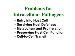
What is an intracellular pathogen? • A microbe that is capable of causing damage to its host that spends at least part of its lifecycle within a host cell or host cell vacuole • ability to replicate within the host cell • ability to persist within the host cell
Why be an intracellular pathogen? • Life outside a cell is harsh • Low pH, shear stress due to circulatory system, complement, antibodies, macrophages, T cells • Bacteria can hide out and replicate inside the cell cytoplasm or inside vacuoles (phagosomes) within cells • Bacteria can invade into underlying tissue
Some notable intracellular pathogens • Coxiella burnetii - Q fever (highly contagious- only a single bacterium required for infection; flu-like symptoms can progress to pneumonia and acute respiratory distress syndrome (ARDS) and death) • Legionella pneumophila* - Legionnaires disease • Listeria monocytogenes* – Listeriosis • Mycobacterium tuberculosis* – Tuberculosis • Rickettsia prowazekii – epidemic typhus (occurs after wars and natural disasters; headache, fever, chills, delirium, death) • Salmonella enterica* – gastroenteritis and typhoid fever • Shigella flexneri – shigellosis (bloody diarrhea and fever, usually resolves within a week)
Entry into a cell • by phagocytosis (cell-eating) • by professional phagocytes like neutrophils, macrophages and dendritic cells • by induced uptake • by cells that are not normally phagocytic (endothelial and epithelial cells)
Phagocytosis Receptor mediated, actin-driven process whereby foreign objects larger than 0.5 mm are internalized and degraded. http://www.nature.com/nri/journal/v8/n2/full/nri2240.html
Phagocytosis animation http://highered.mcgraw-hill.com/sites/0072495855/student_view0/chapter2/animation__phagocytosis.html
Induced uptake Knodler, LA et al 2001 Nat Rev Mol Cell Biol 2(8): 578-588
Host actin • primary host determinant of cell shape and cytoplasmic structure • Rho family of GTPases (Rho, Rac, Cdc42) is important for actin organization • bacteria manipulate the actin cytoskeleton to induce or prevent phagocytosis
Actin cytoskeleton http://scienceblogs.com/transcript/2007/01/cytoskeleton.php
Antiphagocytic action of Y. pestis Yop proteins Cornelis, GR 2002 Nature Rev Mol Cell Biol 3:742-52
Persistence • Two possible mechanisms allowing pathogens to persist within the host cell: • Modification of endocytic pathway • Salmonella enterica • Mycobacterium tuberculosis • Legionella pneumophila • Escape from endocytic vesicle (phagosome) • Listeria monocytogenes • Maybe M. tuberculosis? • Maybe Staphylococcus aureus?
Endocytic and exocytic pathways http://www.landesbioscience.com/curie/chapter/4274/
Endocytic (phagocytic) pathway Haas, A 2007 Traffic 8:311-330
Endocytic (phagocytic) pathway Haas, A 2007 Traffic 8:311-330
Phagosomal Maturation • phagosomes mature due to sequential addition of cellular proteins delivered to them by fusion with different endosomal compartments • anything left in the phagosomes will be destroyed • bacteria need to either • escape before the phagosome environment becomes bactericidal • modify the phagosomal maturation process
Bacteria inside phagosomes -membrane is derived from host plasma membrane -single or multiple bacteria per vesicle -ultimate goal: destruction of bacteria by antimicrobial compounds Haas, A 2007 Traffic 8:311-330
How phagosomes kill bacteria -nicotinamide adenine dinucleotide phosphate (NADPH) oxidase complex produces superoxide radicals from oxygen -nitric oxide synthetase produces NO radicals -vacuolar ATPase pumps protons into the phagosome in reduce pH to ~4.5 -fusion with lysosomes results in delivery of various hydrolases that digest most biological macromolecules Haas, A 2007 Traffic 8:311-330
Ways to study bacterial-phagosomal interactions • Direct microscopic observation • Characterize the vacuoles that contain bacteria using immunofluorescence colocalization • correlate the presence and absence of various host markers with the presence of the bacterium • Directly measure physiological parameters of the phagosome • pH • hydrolytic activities
Colocalization of V-ATPase with bacterial DNA -Monolayer of epithelial cells infected with S. aureus at MOI 10:1 -V-ATPase labelled with antibody -DNA stained with To-Pro-3 Jarry, TM and Cheung, AL 2006 Infect Immun 74 (5):2568-2577
Acidification of a phagosome -measurement of pH following internalization of a bead -concanamycin A inhibits V-ATPase Rohde, K et al 2007 Immunol Rev 219:37-54
Salmonella enterica • Gram negative pathogen • gastroenteritis (self-limiting, mild) to typhoid fever (fatal) • Virulence factors mainly located on one of two Salmonella pathogenicity islands, SPI-1 or SPI-2 • encode type III secretion systems important for invasion of non-phagocytic cells and bacterial replication in phagosomes
Pathogenicity Islands • Large genomic regions that are present in pathogens but absent in their non-pathogenic counterparts • % GC content quite different from surrounding DNA (suggests that DNA acquired from different organism) • Found in both gram negative and gram positive bacteria • Carry virulence genes • Carry factors that promote mobility (integrases, transposases, IS elements)
SPI-1 and SPI-2 Hansen-Wester and Hensel 2001 Microbes Infect 3: 549-559
Intracellular S. enterica Step I: using SPI-1 type III secretion system -injection of effectors that mediate uptake Step II: SPI-2 effectors mediate formation of Salmonella-containing vesicles (SCVs) Knodler, LA et al 2001 Nat Rev Mol Cell Biol 2(8): 578-588
Salmonella-containing vesicle (SCV) • can persist for hours to days • formation requires genes from SPI-2 • characterized by several endosomal markers • EEA1 (early endosomal marker) • Rab5 and Rab11 (middle) • Rab 7, LAMP1, LAMP2, LAMP3, V-ATPase (late) • low pH (indicative of lysosomal fusion) • transient? Steele-Mortimer,Finlay Cell. Micro. (1999) 1(1), 33-49
Salmonella-induced filaments (Sifs) • 4-6 hours after SCV formation, see salmonella-induced filaments (sifs) • not sure what their function in pathogenesis is • not sure what effectors do • Sif formation not yet shown in vivo Rajashekar R et al 2008 Traffic
Salmonella-induced filaments (Sifs) • 4-6 hours after SCV formation, see salmonella-induced filaments (sifs) • not sure what their function in pathogenesis is • not sure what effectors do • Sif formation not yet shown in vivo Rajashekar R et al 2008 Traffic
Salmonella genes important for intracellular life Modulation of host cytoskeleton (SPI-1) SopB-activates Cdc42 and Rho SopE-activates Cdc42, Rac and Rho SopE2-activates Cdc42, Rac and Rho SipA-actin nucleation SipC-actin nucleation Formation and Maintenance of SCV (SPI-2) SifA-Sif formation SopD2- Sif formation SseJ- maintains SCV integrity Steele-Mortimer,Finlay Cell. Micro. (1999) 1(1), 33-49 Haraga A et al 2008 Nat Rev Microbiol 6: 53-66
Mycobacterium tuberculosis • Gram positive • Reservoir: humans • Causes tuberculosis, spread by aerosols • Enters host macrophages and subverts normal phagosome maturation • Persists in a granuloma http://www.sunysb.edu/icbdd/images/infectious2.jpg
Infected macrophages and the granuloma Intracellular M. tuberculosis Granuloma Russell 2007 Nat Rev Microbiol 5:39-47
M. tuberculosis Phagosome • M. tuberculosis • Blocks rab conversion • Retains characteristics of early endosome (Rab5 and EEA-1) • Phagosome pH only reaches pH 6.5 Adapted from Via and Deretic 1997 JBC272:13326-13331
Rab conversion block -Rab5 (early) and Rab7 (late) are GTPases involved in endosome maturation -Rab conversion occurs when Rab5 is replaced with Rab7 Deretic, V. et al 2006 Cell Microbiol 8:719-727
Rab conversion block Mycobacterial factors that cause the block: • Liparabinomannan (LAM) is a lipid (part of mycobacterial cell wall) that prevents the increase of cytosolic [Ca2+] that normally accompanies phagocytosis (and this affects the recruitment of a Rab5 effector) • Phosphatidylinositol mannoside (PIM)- mechanism unknown • SapM is a PI3P phosphatase * *PI3P is important for proper membrane trafficking through the endosomal pathway
Recent observations suggest that M. tuberculosis can escape into the cytosol Direct microscopic observations suggested that M. tuberculosis was not blocking phagosomal maturation at early stages…. -M. tuberculosis in dendritic cells at 2 hours -bacteria (star) colocalize with LAMP-1 (dot)(late endosomal marker) -lysosomes fuse with M. tuberculosis-containing phagosome Van der Wel et al 2007 Cell 129:1287-98
Evidence of escape into cytosol 4 hours -green= bacteria -red= LAMP-1 -blue=host cell DNA -yellow=green and red merged 96 hours *also counted number of bacterial cells at 4 hours and 96 hours and saw increase Van der Wel et al 2007 Cell 129:1287-98
More evidence for escape into cytosol -no phagosomal membrane surrounding M. tuberculosis at 96 hours and no association with LAMP-1 (dots) -escape into cytosol requires RD1 (regions of difference between pathogenic and non-pathogenic mycobacteria) genes (contains ESAT/type VII secretion system) Van der Wel et al 2007 Cell 129:1287-98
Non-pathogenic mycobacterium does not escape into cytosol • -M. bovis (vaccine strain) (green) co-colocalizes (yellow) with LAMP-1 (red) • host cell DNA (blue) -evidence of phagosomal membrane surrounding bacteria -association with LAMP-1 (dots) Van der Wel et al 2007 Cell 129:1287-98
Listeria monocytogenes • Gram positive • Ubiquitous in the environment • Intracellular pathogen • Causes listeriosis • acquired by ingesting contaminated food • mainly affects the elderly, the very young, immunocompromised individuals and pregnant women • results in gastoentiritis, meningitis, encephalitis, septicaemia and death in 25-30% of cases http://www.rps.psu.edu/indepth/graphics/knabel2.jpg
Genes required for L. monocytogenes to invade and spread L. monocytogenes avoids destruction by escaping into host cytosol http://www.sciencedirect.com/science?_ob=ArticleURL&_udi=B6VPN-4NNN0J0-6&_user=1067359&_rdoc=1&_fmt=&_orig=search&_ sort=d&view=c&_version=1&_urlVersion=0&_userid=1067359&md5=30ef1ede32268634f516a8778f263c32
Genes required for survival in the host • inlA and inlB (invasion-associated surface proteins internalin A and B: mediate invasion of non-phagocytic cells) • hly (listeriolysin O or LLO: pore-forming toxin) • plcA and plcB (phospholipase C enzymes: phagocytic vacuole disruption) • actA (actin polymerization) • mpl (metalloprotease required for PlcB maturation) • hpt (hexose phosphate transporter important for rapid growth in host cytosol)
LLO • a primary virulence factor facilitating escape from the phagosome and cell-to-cell spread • member of pore-forming cholesterol-dependent cytolysin family (C. perfringens, S. pyogenes, B. anthracis) • active at pH 5.5 (therefore active in phagosome and not in cytosol)
Mechanism of cholesterol-dependent cytolysin pore formation Monomers bind cholesterol, diffuse laterally…. Tweten 2005 Infect Immun 73:6199-6209
Mechanism of cholesterol-dependent cytolysin pore formation …to form membrane prepore of sufficient size a-helical bundles in prepore are converted into amphipathic transmembrane b-hairpins… Tweten 2005 Infect Immun 73:6199-6209
Mechanism of cholesterol-dependent cytolysin pore formation …and the prepore collapses 40Å such that specific domains are inserted into the membrane The end result is the formation of a large b-barrel pore Tweten 2005 Infect Immun 73:6199-6209
ActA • identified in a screen for bacteria deficient in cell to cell spreading • required for actin-based motility • bacterial surface protein that interacts with several host proteins
Actin comet tails • after a few hours of growth, host actin filaments form on surface of bacteria • polarization into a comet tail (cross-linked network of actin filaments) Cameron LA et al 2000 Nat Rev Mol Cell Biol 1:110-119