Download
1 / 26
310 likes | 552 Views
Respiratory Tract Infection. Upper Respiratory Tract Infection. Lower Respiratory Tract Infection . Lower Respiratory tract Infection. Dr Ali Somily. Objectives. To know the epidemiology and main causes of lower respiratory tract infections

E N D
Respiratory Tract Infection Upper Respiratory Tract Infection Lower Respiratory Tract Infection Lower Respiratory tract Infection Dr Ali Somily
Objectives • To know the epidemiology and main causes of lower respiratory tract infections • The understated the clinical presentation of lower respiratory tract infection • To learn how to diagnose and treat lower respiratory tract infections
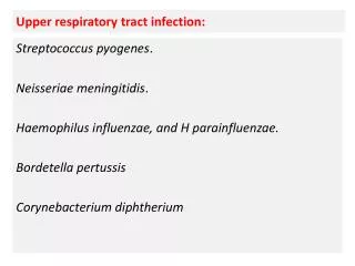
Causes of Respiratory TractInfections • Bacteria • Viruses • Fungi (mainly in the immunocompromised) • Protozoa (in the immunocompromised
Community Acquired Pneumonia • Occurs most frequently in the very young and the very old • May be caused by “typical” or “atypical” pathogens • May secondary to a viral respiratory tract infection
Hospital Acquired Pneumonia • Has the highest mortality • Predisposing factors include abnormal conscious state, intubation, ventilation, surgery and immunosuppression • Is frequently caused by Gram negative organisms
Causes of primary communityacquiredpneumonia Typical pathogens Atypical bacteria • S.pneumoniae • H.influenzae • M.catarrhalis • Staphlococcusaureus (post influenza) • M.pneumoniae (most common) • Chlamydia spp. • Legionaella spp. • Coxiella sp. • M.tuberculosis • Viruses • Influenza A and B • Parainfluenzae • Adenoviruses • Respiratory Syncytial Virus
“Typical” community-acquiredpneumonia • Classically presents with sudden onset of chills, followed by fever, pleuritic chest pain and productive cough. • White blood cell count usually ↑↑ • Sputum, thick, purulent and sometimes rusty coloured. • Chest x-rays show parenchymalinvolvement. • S.pneumoniaeis the commonest cause.
Atypical pneumonia • Various definitions, e.g. • Pneumonia not due to Streptococcus pneumoniae • Pneumonia not responding to beta-lactam therapy • Atypical pneumonia may be primary or • secondary
Atypical pneumonia • Usually, but not always, insidious onset. • Classically patients have non-productive cough, fever, headache and a chest x-ray more abnormal than suggested by clinical examination. • Infection may be sub-clinical and resemble a non-specific viral infection. • M.pneumoniae is the most common organism
Pneumonia complications • Pleural effusion 3-5%: clear fluid +- pus cells • Empyemathoracis 1%: pus in the pleural space (-loculated) • Lung abscess: suppuration + destruction of lung parenchyma • single (aspiration) anaerobes, Pseudomonas • multiple (metastatic) Staphylococcus aureus
15% of Hospital acquired infection • mortality 20-50% • Immuno-compromised patients • General anaesthesia, intubation and ventilation predispose to infection respiratory tract • Gram-negatives, more resistant organisms e.g. Pseudomonas aeruginosa, Enterobacter.
Diagnosis of pneumonia • History and clinical examination • Chest x-ray • Hb, WBC, platelets U+Es, LFTs, ESR • Blood cultures • Sputum – microscopy, culture + sensitivity + virus isolation • Serodiagnosis / antigen detection – if atypical pneumonia suspected
Specimens for microbiology: • Sputum • Contamination with upper respiratory tract/oral flora • Invasive techniques for respiratory samples • Bronchoscopiclavage • Transtrachealaspirate, percutaneousneedle aspiration, open lung biopsy • Non-culture specimens e.g urine for Legionella antigen and pneumococcal antigen in severe disease and PCR for Mycobacterium tuberculosis
Sputum Specimen collection • Teeth brush • Mouthwash • Early morning • Induced sputum if needed • Sterile wide-mouth jar
Therapy of CommunityAcquired pneumonia • In practice it is often not easy to differentiate clinically between “typical” and “atypical” pneumonias. • “Blind therapy” of severe community-acquired pneumonia therefore usually includes a beta-lactamagent + a macrolide. • If the patient is immunocompromisedPneumocystiscarinii must be considered. • The above does not cover viral pneumonia
Therapy of CommunityAcquired Pneumonia (cont’) • Beta-lactams are sometimes given as • monotherapy e.g. amoxycillin. benzylpenicillin, • cefuroxime, cefotaxime, ceftriaxone but • They are inactive against M.pneumoniaeas this does not have a cell wall and have poor activity against intracellular organisms such as Legionellaspp. and Chlamydia spp.
Therapy of pneumonia (cont’) • Erythromycin + rifampicin or a quinolone as mono or combination therapy treatment can be used for pneumonia due to Legionellapneumophilia • Erythromycin or tetracycline or a quinolonecan be used for pneumonia due to Chlamydia spp. or Mycoplasmapneumoniae
Mycoplasmapneumoniae • Acquired by droplet transmission. Epidemics occur every 3-4 years. Occurs in school age children and young adults. • Classically presents with fever, headache, myalgia, earache, mild pharyngitis, dry cough and sometimes arthritis. • Skin rashes and haemolyticanaemiamay occur. • Neurological complications occasionally happen.
Diagnosis of M.pneumoniae • Clinical and CXR shows a patchy bilateral bronchopneumonia • Culture fried egg colony (M.homonis) • Cold agglutinins may be present • IgM test • Serology
Legionellosis • Caused by Legionellapneumophilia. Serogroup 1 • L.pneumophiliamay cause a multi-system disease with confusion, muscle aches, pneumonia, renal failure, liver involvement + diarrhoea and significant mortality. • L.pneumophiliamay also cause Pontiac fever – a self-limiting disease.
Legionellosis diagnosis(general) • History (exposure to cooling towers, etc) and clinical. • Laboratory • Culture on buffered charcoal-yeast extract (BCYE) agar , serology or urine antigen detection • CXR Patchy interstitial involvement or consolidation • Hyponatraemia often present • Urea frequently raised • Liver function test is abnormal
Therapy for Pneumonia caused by LegionellaPneumophilia • In severe legionella pneumonia traditionally a combination of a parenteralmacrolideand rifampicinis used • Fluoroquinolones may be better than macrolides
Chlamydia pneumoniae • Person-to-person spread occurs • Causes atypical pneumonia • Implicated as potential pathogen / co-pathogen in coronary artery disease and cerebrovasculardisease • Diagnosis by immunofluorescence, cell culture using McCoy cell or serology
Chlamydia psittaci • Is a zoonosis acquired from birds • Human disease acquired by inhalation of infected aerosols • Causes atypical pneumonia • Diagnosis is usually by serology .
Q fever • Caused by Coxiellaburnetii • Transmitted via infected animals through milk, excreta, etc. • Can cause atypical pneumonia. • Diagnosis usually by serology
Exacerbations of ChronicObstructive Pulmonary Disease • May be due to infection – viral or bacterial • Implicated bacteria include S.pneumoniae, H. influenzae. M. catarrhalis and coliforms • M. catarrhalis almost always produces betalactamaseand so will not repond to amoxicillin therapy