Download
1 / 13
130 likes | 357 Views
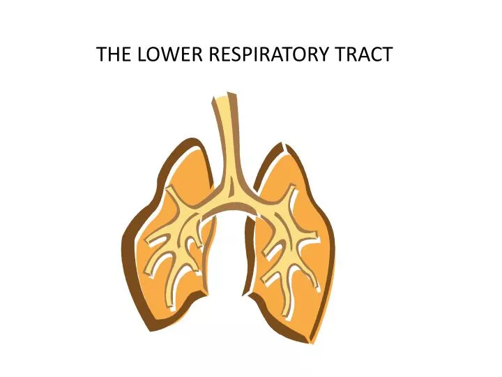
THE LOWER RESPIRATORY TRACT. OFTEN REFERRED TO AS THE TRACHEOBRONCHIAL TREE AFTER AIR LEAVES LARYNX AND ENTERS THE TRACHEA ALSO CALLED WINDPIPE 4 ½ “ LONG IT IS LINED WITH CILIATED MUCOUS MEMBRANE WE REMOVE DUST OR MUCUS FROM THE TRACHEA BY COUGHING OR EXPECTORATING

E N D
OFTEN REFERRED TO AS THE TRACHEOBRONCHIAL TREE • AFTER AIR LEAVES LARYNX AND ENTERS THE TRACHEA • ALSO CALLED WINDPIPE • 4 ½ “ LONG IT IS LINED WITH CILIATED MUCOUS MEMBRANE • WE REMOVE DUST OR MUCUS FROM THE TRACHEA BY COUGHING OR EXPECTORATING • DEEP BREATHS AND THEN A FORCEFUL EXPULSION OF AIR • EXTENDS FROM CRICOID CARTILAGE OF LARYNX TO 6TH THORACIC VERTEBRAE • C- CARTILAGE • PROTECTION • ALLOWS ESOPHAGUS TO EXPAND WITH SWALLOWING
Bifurcation – branching • Trachea bifurcates in the center of the chest into 2 bronchi • Right mainstem and left mainstem • Primary bronchi • Carina – site of bifurcation • The bronchi branch again into the 5 lobar bronchi • Correspond to the 5 lobes of the lung • The lobar bronchi branch into the segmental bronchi
Tissue layers of bronchi are the same from the trachea to the segmental bronchi • First layer is epithelial tissue- contains mucociliaryescaltor (pseudostratified ciliated columnar cells) • Keeps area clean of debris • Middle layer – smooth muscle , lymph, & nerve tracts • Third layer is the protective layer- cartiliginous • Segmental bronchi divide into subsegmental bronchi • Branch deep into each lung segment • Cartiliginous c-rings begin to disappear
Bronchioles branch off subsegmental bronchi • No cartilage • Epithelial lining now is ciliated cuboidal cells • Goblet cells, cilia , submucosal glands are almost gone • Still just conduction of air – no gas exchange yet • Terminal bronchioles branch off the bronchioles • No cilia, goblet cells, cartilage, submucosal glands • THE END OF THE CONDUCTING AREA ( movement of air)
Respiratory bronchiole branches off the terminal bronchiole • A small portion of gas exchange takes place here • Simple cuboidal cells in between true alveoli cells • Pancake like cells called simple squamous pneumocytes • Alveolar ducts originate from here • Simple squamous cells arrange in a tube-like pattern • Ducts give way to the alveoli- also known as alveolar ducts
Alveolar capillary membrane • Alveoli are surrounded by numerous pulmonary capillaries • 300-600 million alveoli in adult lung • About the size of a tennis court is available for diffusion of O2 & CO2 • Blood in pulmonary capillaries is rich in CO2 • it just came from the right side of the heart • External respiration takes place here • CO2 leaves blood and O2 enters blood
Layers of the alveolar capillary membrane • first layer – is liquid surfactant which lines the alveoli • Helps lower the surface tension in the alveoli • No surfactant = collapsed alveoli • Second layer – alveolar epithelium • 2 different cells • Type I – squamous pneumocyte • Gas molecules easily pass through process of gas exchange • Type II – granular pneumocytes come in • Produce surfactant • Aid in cellular repair
Type III cells • Wandering macrophages- clean up the area so gas exchange can take place • Kohn pores • Small holes between alveoli so gases and macrophages can move between • Third layer of alveolar capillary membrane • Interstitial space • Space between the basement layer of the alveoli and the basement layer of the capillary • Appears fused because it is so small • Interstitial edema – fluid in this space • Membranes separate and gas exchange becomes harder • Fourth layer • Capillary endothelium(simple squamous epithelium) • Forms wall of capillary