Download
1 / 6
60 likes | 175 Views
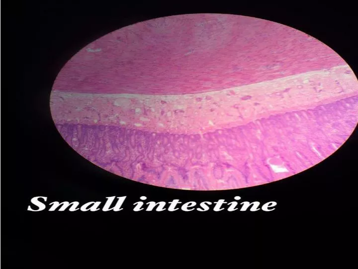
Simple columnar epithelium “villi” Submucosa ; Forms core of Plica circularis . Muscularis externa. Villi clubbed shape. Lamina Propria ( Peyer's patches). Villi leaf shape Submucosa ; Duodenal (Brunner ’ s)Gland s. Simple columnar epithelium Tenia coli . No villi Lymph nodules

E N D
Simple columnar epithelium “villi” • Submucosa; • Forms core of Plicacircularis. • Muscularisexterna
Villi clubbed shape. • Lamina Propria(Peyer's patches).
Villi leaf shape Submucosa; Duodenal (Brunner’s)Glands.
Simple columnar epithelium Tenia coli
No villi Lymph nodules Mesoappendix


![Falling in Love with Forms [BlendConf 2014]](https://cdn4.slideserve.com/7566422/falling-in-love-with-forms-dt.jpg)
![Falling in Love With Forms [Breaking Development Nashville 2015]](https://cdn4.slideserve.com/7566897/falling-in-love-with-forms-dt.jpg)
![Falling in Love with Forms [Øredev 2015]](https://cdn4.slideserve.com/7567052/falling-in-love-with-forms-dt.jpg)
![The Features of Highly Effective Forms [SmashingConf NYC 2016]](https://cdn4.slideserve.com/7568502/the-features-of-highly-effective-forms-dt.jpg)