Download
1 / 25
250 likes | 478 Views
Mammalian cell genetics Introduction: Genetics as a subject (genetic processes that go on in somatic cells: that replicate, transmit, recombine, and express genes) Genetics as a tool . Most useful the less you know about a process. 4 manipulations of genetics: 1- Mutation :

E N D
Mammalian cell genetics Introduction: Genetics as a subject (genetic processes that go on in somatic cells: that replicate, transmit, recombine, and express genes) Genetics as a tool. Most useful the less you know about a process. 4 manipulations of genetics: 1- Mutation: in vivo (chance + selection, usually); targeted gene knock-out or alteration in vitro: site directed or random cassette 2- Mapping: Organismic mating segregation, recombination (e.g., transgenic mice); Cell culture: cell fusion + segregation; radiation hybrids; FISH 3- Gene juxtaposition (complementation): Organisms: matings phenotypes of heterozygotes; Cell culture: cell fusion heterokaryons or hybrid cells 4- Gene transfer: transfection Last updated Nov. 16, 12:10 AM
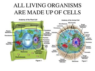
Mammalian cell genetics Advantages of cultured cells (vs. whole organism): numbers, homogeneity Disadvantages of cultured mammalian cells: limited phenotypes limited differentiation in culture (but some phenotypes available) no sex (cf. yeast) Mammalian cell lines(previously discussed) Most genetic manipulations use permanent lines, for the ability to do multiple clonings Primary, secondary cultures, passages, senescence. Crisis, established cell lines, immortality vs. unregulated growth. Most permanent lines = immortalized, plus "transformed“, (plus have abnormal karyotypes)
Mutation in cultured mammalian cells: Problem of epigenetic change: variants vs. mutants Variants could be due to: Stable heritable alterations in phenotype that are not due to mutations: heritable switches in gene regulation (we don’t yet understand this). DNA CpG methylation Chromatin organization: e.g., histone acetylation (active) / de-acetylation (inactive) Diploidy. Heteroploidy. Haploidy. The problem of diploidy and heteroploidy: Recessive mutations (most knock-outs) are masked. (cf. e.g., yeast, or C. elegans, Dros., mice): f2 homozygotes)
Solutions to diploidy problem: Double mutants: heavy mutagenesis, mutants/survivor increases but mutants/ml decreases Incl. also mutation + segregation, or mutation + homozygosis: (rare but does occur) How hard is it to get mutants? What are the spontaneous and induced mutation rates? (loss of function mutants) Spont: ~ 10-7/cell-generation Induced: ~ 2 x 10⁻⁴ to 10-3/cell(EMS, UV) So double knockout could be 0.00072~ 5X10⁻⁷. One 10cm tissue culture dish holds ~ 107 cells. Note: Same considerations for creation of recessive tumor suppressor genes in cancer: requires a double knockout. But there are lots of cells in a human tissue or in a mouse. RNAi screen, should knock down both alleles: Transfect with a library of cDNA fragments designed to cover all mRNAs. Select for knockout phenotype (may require cleverness). Clone cells and recover and sequence RNAi to identify target gene. A human near-haploid cell strain. Use of it: Science, 326: 1231-1235 (2009) EMS = ethyl methanesulfonate: ethylates guanine UV (260nm): induces dimers between two adjacent pyrimidines on the same DNA strand
L R R L Homozygosis: Loss of heterozygosity (LOH)by mitotic recombination between homologous chromosomes (rare) M i t o s i s - - - + + 2 heterozygotes again L R L R L R R L or - - + + - - + + Paternal Chr. 4, say Maternal Chr. 4 - - Recombinant chromatids + + After homologous recombination (not sister chromatid exchange) Heterozygote 1 homozygote +/+1 homozygote -/- Recessive phenotype is unmasked = a mechanism of homozygosis of recessive tumor suppressor mutations in cancer
Mutagenesis (induced general mutations, not site directed) Chemical and physical agents: MNNG point mutations (single base substitutions) EMS point mutations (single base substitutions) Bleomycin small deletions UV mostly point mutations but also large deletions Ionizing radiation (X-, gamma-rays) large deletions, rearrangements Dominant vs. recessive mutations; Dom. are rare (subtle change in protein), but expression easily observed, Recessives are easier to get (whatever KO’s the protein function), but their expression is masked by the WT allele. MNNG = methyl N-nitrosoguanidine EMS = ethylmethane sulfonate
Categories of cell mutant selections Example • Auxotrophs (via BrdU selection) purine-; pyrimidine-; glyc-, pro-;gln- • Drug resistance Dominant ouabainR, alpha-amanitinR Recessive 6-TGr, BrdUr • Antibodies vs. surface components MHC- • Visual inspection G6PD-, Ig IP- • FACS = fluorescence‑activated cell sorter DHFR- • Brute force screening IgG-, electrophoretic shifts • Temperature‑sensitive mutants 3H-leu resistant (leucyl tRNA synthetase-) TG = 6-thioguanine; BrdU = 5-bromodeoxyuridine; MHC = majpor histocompatibility locus; G6PD = glucose-6-phosphate dehydrogenase
Auxotroph selection by killing growing cells: Mutant cell cannot grow in deficient medium so does not incorporate BrdU (BUdR) and so survives DNA damage from subsequent treatment with 313 nm light Kao and Puck, PNAS
Purine biosynthesis, salvage pathways, and inhibitors Adenine(A) (diaminopurine) (8-azaadenine) Methotrexate Folate (=amethopterin) (~aminopterin) Adenosine APRT Adenosine FH4 kinase Nuc. Acid AMP Glycine Thymidine (T) Adenylosucc. Alanosine PRPP + IMP glutamine HGPRT Azaserine XMP GMP Nuc. Acid Hypoxanthine Glutamine XGPRT HGPRT (H) (Eco gpt) Guanine Xanthine Mycophenolic (6-thioguanine) (X) acid (8-azaguanine) Salvage enzymes Biosynthesis; Analogs (iytal.) Code: Inhibitors DHFR (drugs, in italics) PRPP = phosphoribosyl pyrophosphate; FH4=tetrahydrofolate
Purine biosynthesis, salvage pathways, and inhibitors Adenine(A) (diaminopurine, DAP) (8-azaadenine, 8AA) Methotrexate Folate (=amethopterin) (~aminopterin) Adenosine APRT Adenosine FH4 kinase Nuc. Acid AMP Glycine Thymidine (T) Adenylosucc. Alanosine PRPP + IMP glutamine HGPRT Azaserine XMP GMP Nuc. Acid Hypoxanthine Glutamine XGPRT HGPRT (H) (Eco gpt) Guanine Xanthine Mycophenolic (6-thioguanine, 6TG) (X) (8-azaguanine, 8AG) Test yourself: Fill in the boxes Grow (+) or not grow(-) Click here for the answers Growth pattern examples GHT = glycine, hypoxanthine, and thymidine A = adenine H = hypoxanthine G = glycine TG = 6-thioguanine (G analog) DAP = diaminopurine (A analog) MTX = methotrexate (DHFR inhibitor) DHFR = dihydrofolate reductase HPRT = hypoxanthine-guanine phosphoribosyltransferase APRT = adenine phosphoribosyltransferase in italics - + + -
Cell mutant types: • 1. Auxotrophs (BrdU reverse selection) • 2. Drug resistance (dominants or recessives) • 3. Temperature‑sensitive mutants: cell cycle mutants. • Tritiated amino acid suicide (aa‑tRNA synthetases) • 4. Antibody resistance. Lysis with complement. Targets cell surface constituents mostly (e.g., MHC) • 5. Visual inspection at colony level: • A. Sib selection (G6PD) • B. Replica plating (LDH) • C. Secreted product (Ig: anti-Ig IP) • FACS = fluorescence‑activated cell sorter (cell surface antigen or internal ligand binding protein) • Brute force (clonal biochemical analysis, e.g., electrophoretic variants (e.g., Ig, isozymes)) • MHC = major histocompatability locus or proteins • G6PD = glucose-6-phosphate dehydrogenase; • LCH = lactate dehydrogenase; Ig = immunoglobulin. IP = immunoprecipitate
Cell fusion (for gene juxtaposition, mapping, protein trafficking, etc. ) Fusogenic agents PEG, Sendai virus (syncytia promoting, as HIV). Heterokaryons (2 nuclei), no cell reproduction (limited duration). (e.g., studied membrane fluidity, nuclear shuttling, gene activation (myoblasts)) Hybrids (nuclei fuse, some cells (minority) survive and reproduce). Small % of heterokaryons. Complementation (e.g., auxotrophs with same requirement) allows selection Dominance vs. recessiveness can be tested. Chromosome loss from hybrids Mapping: chromosome assignment synteny. Radiation hybrids: linkage analysis (sub-chromosomal regional assignments). PEG =polyethylene glycol, (available 1000 to 6000 MW)
Cell fusion Hprt+, TK- + Parental cells Hprt-, TK+ HAT- HAT- PEG (polyethylene glycol, mw ~ 6000 Sendai virus, inactivated Cell fusion Heterokaryon (alternative = a homokaryon) HAT medium Hprt-, TK+ Hprt+ TK- Hybrid cell HAT+ Cell cycle, Nuclear fusion, Mitosis, Survival, reproducton Hprt-, TK+, Hprt+ TK- Heterokaryon use examples: membrane dynamics (lateral diffusion of membrane proteins) shuttling proteins (e.g., hnRNP A1 ), gene regulation (e.g., turn on myogenesis) Hybrid cells: examples of use: gene mapping (synteny) gene regulation (dominance/recessiveness) Complementation Synteny = genes physically linked on the same chromosome are syntenic.
Frye and Edidin, 1970: Use of cell fusion and heterokaryons to measure the diffusion of membrane proteins t=0 t=40’ Complete mixing in < 40 min. No diffusion at low temperature (<15-20 deg) http://www.erin.utoronto.ca/~w3bio315/lecture2.htm
Complementation analysis Mutant parent 1 Mutant parent 2 Mutant parent 1 Mutant parent 2 gly2- + + gly3- gly1- gly1- Cell fusion Cell fusion Hybrid cell Hybrid cell glyA- glyA- glyA- glyB- Glycine-free medium: No growth No complementation same gene (named glyA) Glycine-free medium: Yes, growth Yes, complementation different genes genes (named glyA and glyB)
Nuclear-cytoplasmic shuttling demonstrated using interspecific heterokaryons Unfused frog cells HnRNP A1 HnRNP C Fused cell: HeLa + frog A1 shuttles, C does not. Pinal-Roma and Dreyfuss, Nature, 355:730 Frog nuclei in fused cell CHX = cycloheximide (protein synthesis inhibitor) given 0.5 h before fusion
DNA polybrene DNA Linear PEI DNA transfection Transfection agents: CaPO4 (co-precipitates with DNA) Electroporation (naked DNA, high voltage pulse transient holes) Lipofection (multilamellar liposomes) Polybrene (detergent) Ballistic (DNA-coated gold particles) DEAE-dextran (toxic, OK for transient) Poly-ethylenimine (PEI, cheap) Effectene (non-liposomal lipid) Must traverse cytoplasm. Much engulfed in lysosomes. Inhibition of lysosomal function often helps (chloroquin). Co-integration of high MW DNA . Can = 2000 KB. Separate plasmids transfected together same site (co-integration). Separate transfections separate locations Random or semi‑random (many) integration sites (unless targeted) Low but real homologous recombination rate. History: mammalian cell transfection developed for practical use at Columbia (at P&S: Wigler, Axel and Silverstein) DEAE= diethyl-amino-ethyl (positively charged)
Mike Wigler Richard Axel Saul Silverstein History: discovered for practical use at Columbia (P&S: Wigler Axel and Silverstein)
Transient transfection vs. permanent: cloned genes Unintegrated DNA chromosomally integrated. Unnatural? Position effects ? Super-physiological expression (so analyze a pool of many to levels (per transfected cell) ? average) Transient -> 10‑90% transfection efficiency (stain) Permanents more like 0.001 transfectants per μg DNA per cell (~high). i.e., 106 treated cells -> 1000 colonies; could be much less for certain types of cells
One the most dramatic first applications of gene transfection from total DNA: Transfer of the growth‑transformed phenotype: ability to grow in multilayers or in suspension in soft agar: (Weinberg; Wigler) DNA from tumor transfected into growth-controlled mouse 3T3 cells. Look for foci (one = focus). Make a library from growth‑transformed transfectant. Screen for human Alu repeat. Verify that cloned DNA yields high frequency of focus‑forming transfectants. Isolate cDNA by hybridization to the cloned genomic DNA. Sequence. Identify gene: = a dominant oncogene. Ras, a signaling protein in a transducing pathway for sensing growth factors Transformed Mouse 3T3 cells transfected with an EGFreceptor gene Mouse 3T3 cells
Recombination; gene targeting Mitotic recombination between homologous chromosomes; relation to cancer through the loss of tumor suppressor genes LOH = loss of homozygosity: WT = +/+ mutation +/- (WT phenotype) (LOH via homologous recombination in G2; or chromosome loss and duplication) -/- (mutant phenotype revealed) Recombination of transfecting genes: homologous (rare) vs. non‑homologous (common) recombination.
Gene knockouts via homologous recombination ES cells and transgenic mice. Selection for homologous recombinants via the loss of HSV TK genes (Capecchi): –tk – homol. region – drugR – homol. region – tk – Non-homologous recombination favors ends; tk is inserted, conferring sensitivity to the drug gancyclovir (HSVtk specific, not a substrate for human tk) Most work in ES cells mice homozygosis via F1 breeding Little work in cultured lines: Myc double sequential K.O. = viable, ~sick (J. Sedivy) Splicing factor (ASF) double K.O. see next graphic. APRT = adenine phosphoribosyltransferase ASF = alternative splicing factor
HSV-TK gene is removed during homologous recombination, but remains joined during non-homologous recombination. Unlike mammalian TK, HSVTk converts gancyclovir to a toxic product HSV = Herpes simplex virus; tk = thymidine kinase; FIAU = equivalent to gancyclovir, today Resistant to gancyclovir Die in gancyclovir M. Capecchi, Nature Medicine7, 1086 - 1090 (2001) Generating mice with targeted mutations
ASF ASF neo ASF ASF ASF Human Human Human Human Human ASF ASF ASF ASF ASF Double knockout of the ASF gene, a vital gene, by homologous recombination Chicken DT40 cells One ASF gene allele disrupted by homologous recombination + hol ASF- hol neo neo Tet-off promoter pur Hol = histidinol resistance; pur = puromycin resistance Drug resistance genes here chosen for illustration. Both alleles have been disrupted in some purR, holR cells neo ASF- neo +tet pur ASF- pur X Wang, Takagaki, and Manley, Targeted disruption of an essential vertebrate gene: ASF/SF2 is required for cell viability. Genes Dev. 1996 Oct 15;10(20):2588-99. Cell dies without ASF(follow events biochemically) cell viable(covered by human ASF gene
NAD+ Histidinol dehydrogenase Histidinol dehydrogenase detoxifies histidinol, confers histidinol resistance protein synthesis inhibits protein synthesis (charged to tRNA but cannot be transferred to growing peptide so truncates)