Download
1 / 48
500 likes | 783 Views
Chapter 9 - Lipids and Membranes. Lipids are essential components of all living organisms Lipids are water insoluble organic compounds They are hydrophobic (nonpolar) or amphipathic (containing both nonpolar and polar regions ). Fig 9.1 Structural relationships of major lipid classes.

E N D
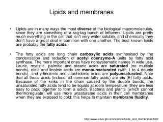
Chapter 9 - Lipids and Membranes • Lipids are essential components of all living organisms • Lipids are water insoluble organic compounds • They are hydrophobic (nonpolar) or amphipathic (containing both nonpolar and polar regions)
Fatty Acids • Fatty acids - R-COOH (R=hydrocarbon chain) are components of triacylglycerols, glycerophospholipids, sphingolipids • Fatty acids differ from one another in: • (1) Length of the hydrocarbon tails • (2) Degree of unsaturation (double bond) • (3) Position of the double bonds in the chain Nomenclature of fatty acids • Most fatty acids have 12 to 20 carbons • Most chains have an evennumber of carbons • IUPAC nomenclature: carboxyl carbon is C-1 • Common nomenclature:a,b,g,d,eetc. from C-1 • Carbon farthest from carboxyl is w
Structure and nomenclature of fatty acids • Saturated - no C-C double bonds • Unsaturated - at least one C-C double bond • Monounsaturated - only one C-C double bond • Polyunsaturated - two or more C-C double bonds Double bonds in fatty acids • Double bonds are generally cis • Position of double bonds indicated byDn, where n indicates lower numbered carbon of each pair • Shorthand notation example: 20:4D5,8,11,14 • (total # carbons : # double bonds,D double bond positions) Chapter 9
Fig. 9.3 Structures of three C18 fatty acids • (a) Stearate (octadecanoate) • (b) Oleate (cis-D9-octadecenoate) • (c) Linolenate (all-cis-D9,12,15-octadecatrienoate) • The cis double bonds produce kinks in the tails of unsaturated fatty acids
Triacylglycerols • Fatty acids are stored as neutral lipids, triaclyglycerols (TGs)Fats have 2-3 times the energy of proteins or carbohydrates • TGs are 3 fatty acyl residues esterified to glycerol • TGs are hydrophobic, stored in fat cells (adipocytes) Fig 9.5 Structure of a triacylglycerol
Fig 9.10 Structure of an ethanolamine plasmalogen • Plasmalogens - C-1 hydrocarbon substituent attached by a vinylether linkage (not ester linkage)
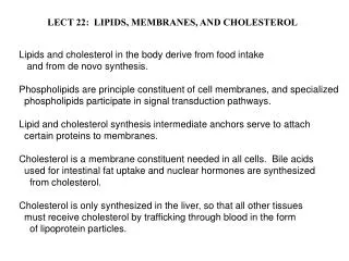
Sphingolipids • Sphingolipids- sphingosine is the backbone abundant in central nervous system tissues • Ceramides - fatty acyl group linked to C-2 of sphingosine by an amide bond • Sphingomyelins - phosphocholine attached to C-1 of ceramide • Cerebrosides- glycosphingolipids with one monosaccharide residue attached via a glycosidic linkage to C-1 of ceramide • Galactosylcerebrosides - a singleb-D-galactose as a polar head group • Gangliosides - contain oligosaccharide chains with N-acetyl-neuraminic acid (NeuNAc) attached to a ceramide Chapter 9
Fig 9.11 (a) Sphingosine (b) Ceramides (c) Sphingomyelin
Fig 9.12 • Structure of a galactocerebroside
Steroids • Classified as isoprenoids - related to 5-carbon isoprene (found in membranes of eukaryotes) • Steroids contain fourfusedring systems: 3-six carbon rings (A,B,C) and a 5-carbon D ring • Ring system is nearly planar • Substituents point either down (a) or up (b) Fig 9.14 Isoprene Fig 9.15 Chapter 9
Cholesterol • Cholesterol modulates the fluidity of mammalian cell membranes • It is also a precursor of the steroid hormones and bile salts • It is a sterol (has hydroxyl group at C-3) • The fused ring system makes cholesterol less flexible than most other lipids
Cholesterol esters • Cholesterol is converted to cholesteryl esters for cell storage or transport in blood • Fatty acid is esterified to C-3 OH of cholesterol • Cholesterol esters are very water insoluble and must be complexed with phospholipids or amphipathic proteins for transport Fig 9.17 Cholesteryl ester
Waxes • Waxes are nonpolar esters of long-chain fatty acids and long chain monohydroxylic alcohols • Waxes are very water insoluble and high melting • They are widely distributed in nature as protective waterproof coatings on leaves, fruits, animal skin, fur, feathers and exoskeletons Fig 9.18 Myricyl palmitate, a wax
Eicosanoids • Eicosanoids are oxygenated derivatives of C20 polyunsaturated fatty acids (e.g. arachidonic acid) • Prostaglandin E2 - can cause constriction of blood vessels • Thromboxane A2 - involved in blood clot formation • Leukotriene D4 - mediator of smooth-muscle contraction and bronchial constriction seen in asthmatics • Aspirin alleviates pain, fever, and inflammation by inhibiting cyclooxygenase (COX), an enzyme critical for the synthesis of prostaglandins. (NSAID family of compounds)
Lipid vitamins • Vitamins A,D,E, and K are isoprenoid derivatives Vitamin E
Biological Membranes Are Composed of Lipid Bilayers and Proteins • Biological membranes define the external boundaries of cells and separate cellular compartments • A biological membrane consists of proteins embedded in or associated with a lipid bilayer Chapter 9
Several important functions of membranes • Some membranes contain proteinpumps for ions or small molecules • Some membranes generate protongradients for ATPproduction • Membrane receptors respond to extracellular signals and communicate them to the cell interior Lipid Bilayers • Lipid bilayers are the structural basis for all biological membranes • Noncovalent interactions among lipid molecules make them flexible and self-sealing • Polar head groups contact aqueous medium • Nonpolar tails point toward the interior
Fluid Mosaic Model of Biological Membranes • Fluid mosaic model - membrane proteins and lipids can rapidly diffuse laterally or rotate within the bilayer (proteins “float” in a lipid-bilayer sea) • Membranes: ~25-50% lipid and 50-75% proteins • Lipids include phospholipids, glycosphingolipids, cholesterol (in some eukaryotes) • Compositions of biological membranes vary considerably among species and cell types
Lipid Bilayers and Membranes Are Dynamic Structures Fig 9.23 (a) Lateral diffusion is very rapid (b) Transverse diffusion (flip-flop) is very slow
Fig 9.25 Freeze-fracture electron microscopy, distribution of membrane proteins
Three Classes of Membrane Proteins • (1) Integral membrane proteins • Contain hydrophobic regions embedded in the lipid bilayer • Usually span the bilayer completely (2) Peripheral membrane proteins • Associated with membrane through charge-charge or hydrogen bonding interactions to integral proteins or membrane lipids • More readily dissociated from membranes than covalently bound proteins • Change in pH or ionic strength often releases these proteins (3) Lipid-anchored membrane proteins • Tethered to membrane through a covalentbond to a lipid
Membrane Transport • Three types of integral membrane protein transport: • (1) Channels and pores • (2) Passive transporters • (3) Active transporters Chapter 9
Pores and Channels • Pores and channels are transmembrane proteins with a centralpassage for ions and small molecules • Solutes of appropriate size, charge, and molecular structure can diffuse down a concentration gradient • Process requires no energy • Central passage allows molecules and ions of certain size, charge and geometry to transverse the membrane. • Figure 9.30
Passive Transport • Passive transport (facilitateddiffusion) does not require an energy source • Protein binds solutes and transports them down a concentration gradient • Uniport - transporter carries only a single type of solute • Some transporters carry out cotransport of two solutes, either in the samedirection (symport) or in oppositedirections (antiport) Types of passive transport systems
Fig 9.31 • Types of passive transport(a) Uniport(b) Symport(c) Antiport
Fig 9.32 Kinetics of passive transport • Initial rate of transport increases until a maximum is reached (site is saturated)
Active Transport • Transport requires energy to move a solute up its concentration gradient • Transport of charged molecules or ions may result in a charge gradient across the membrane • Primary active transport is powered by a direct source of energy as ATP, light or electron transport • Secondary active transport is driven by an ion concentration gradient Types of active transport Chapter 9
Fig 9.34 Secondary active transport in E. coli • Oxidation of Sred generates a transmembrane proton gradient • Movement of H+ down its gradient drives lactose transport (lactose permease)
Fig 9.35 Secondary active transport in animals: Na+-K+ ATPase • Na+ gradient (Na+-K+ATPase) drives glucose transport
Endocytosis and Exocytosis • Cells import/export molecules too large to be transported via pores, channels or proteins by: • Endocytosis- macromolecules are engulfed by plasma membrane and brought into the cell inside a lipid vesicle • Exocytosis - materials to be excreted from the cell are enclosed in vesicles that fuse with the plasma membrane
Transduction of Extracellular Signals • Specific receptors in plasma membranes respond to external chemicals (ligands) that cannot cross the membrane: hormones, neurotransmitters, growth factors • Signal is passed through membrane proteintransducer to a membrane-bound effector enzyme • Effector enzyme generates a second messenger which diffuses to intracellular target
Fig 9.37 General mechanism of signal transduction across a membrane
Fig 9.43 • Summary of the adenyl cyclase signaling pathway