Download
1 / 25

E N D
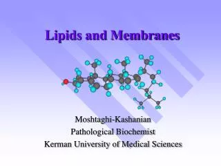
Lipids and membranes • Lipids are in many ways the most diverse of the biological macromolecules, since they are something of a rag-tag bunch of leftovers. Lipids are pretty much everything in the cell that isn't very water soluble, and chemically they don't have a great deal in common with one another. The best known lipids are probably the fatty acids. • The fatty acids are long chain carboxylic acids synthesised by the condensation and reduction of acetyl coenzyme-A units by fatty acid synthase. The more important ones have nonsystematic names in wide use. Lauric, myristic, palmitic and stearic acids are saturated (no multiple bonds), oleic and linoleic acids are monounsaturated (with 1 to 4 double bonds), and γ-linolenic and arachidonic acids are polyunsaturated. Note that all these acids (indeed, all common fatty acids) are cis (E) fatty acids. Because of the kinks in the chain caused by the double bonds, the unsaturated fatty acids tend to be liquids at room temperature (they are less easy to pack together to form a solid). Bacteria and plants (which cannot thermoregulate) will use more unsaturated acids in their cell membranes when they are exposed to cold: this helps to maintain membrane fluidity. http://www.steve.gb.com/science/lipids_and_membranes.html
Complex lipids Triacylglycerols Non polar lipids are storage lipids Phosphatidylcholine Glycerophospholipids are membrane forming lipids
Properties of membrane phospholipids In unsaturated fatty acids, these chains are kinked, and cannot pack so closely. This increases membrane fluidity. • Membrane phospholipids are amphipathic. The head group, containing e.g. charged phosphate and polar choline, is hydrophilic and water soluble. • The tail group, containing a nonpolar fatty acid chain, is hydrophobic (lipophilic) and water insoluble.
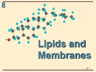
Sterols • In addition to the glycerolipids and sphingolipids, eukaryotes make wide use of sterols and steroids. These hydrophobic compounds are derived from isoprene, and belong to the group of chemicals termed 'terpenes'. Cholesterol is a common sterol component of eukaryotic cell membranes (but is absent from almost all bacteria). It is the precursor for many hormones, too. Cholesterol
Models of membranes: Langmuir's monolayers • Langmuir showed that if phospholipids are dissolved in benzene they could be dispersed as a monolayer on the surface of water in a Langmuir trough. • A monolayer consists of a thin layer of amphipathic molecules arranged with their polar groups dissolved in water, and their nonpolar group dissolved in each other.
Models of membranes: Micelles • If shaken with water, phospholipids (like detergents) will form micelles. These are colloids in an aqueous suspension. Micelles have a hydrophilic outside and hydrophobic inside. • Water is inherently disordered, but when lipids are present, the water molecules have to arrange themselves in a more ordered way. The less order there is in a arrangement, the more likely (∆G = ∆H + T∆S) the arrangement is to happen. The clumping together of fat molecules reduces the amount of water that has to be ordered. The left hand (clumpy) picture has fewer water molecules arranged neatly than the right hand (less clumpy) picture
Models of membranes: Bilayer • Gortner & Grendel's bilayers (1925) • These researchers extracted the lipid from the plasma membrane of red blood cells and applied them to a Langmuir trough. They covered twice the area of the original membrane showing that natural membranes are bilayers.
Models of membranes: Liposomes • If lipids are sonicated at 20 kHz they form vesicles (liposomes) with an internal space. These can be used to deliver hydrophobic drugs to cells, and as model cells.
Membrane Models • Davison & Danielli's sandwich (1935) • The earliest true model of membranes proposed a phospholipid bilayer covered in a globular protein coat. • Singer-Nicholson fluid mosaic (1972) • The fluid mosaic model pictures the membrane as a phospholipid bilayer with many proteins, some integral to the membrane, others attached more loosely. Note the many other components, such as cholesterol; and the attachement sites for the extracellular environment (via glycoproteins) and intracellular cytoskeleton.
Membrane proteins • Peripheral membrane proteins attach via non-covalent interaction with other membrane proteins. They can be removed easily with gentle persuasion (e.g. high ionic strength buffers) • In integral membrane proteins hydrophobic amino acids form an α-helix (with the hydrophobic residues pointing outward into the inside of the membrane), which spans the membrane. Carbohydrates coat the hydrophilic extra-cellular portions, and such proteins are termed glyco-proteins. They are difficult to remove from membranes with gentle procedures. • Pore proteins are integral proteins in which hydrophobic amino acids line the outside of the pore, binding the pore into the membrane, whilst hydrophilic amino acids line the inside of the pore, providing a channel that is friendly to water-soluble chemi-cals.
Mobility in a membrane • Fatty acids and proteins are able to diffuse in the membrane, either laterally (parallel to the membrane), or transversely (flip-flopping from one leaflet of the membrane to the other). Lateral diffusion of phospholipids is far faster than transverse 'flip-flop'. Proteins can also migrate laterally. If we fuse a mouse and a human cell, the membrane proteins are thoroughly mixed within 1 hour.
Transport across membranes Membranes are (approximately) thin layers of lipids, and consequently their cores are very hydrophobic. There question evolves, how water-soluble components are transported across these membranes Chemicals can cross in three main ways. • Unmediated (simple) diffusion: chemical species cross the hydrophobic core by simple diffusion down a chemical gradient. • Facilitated diffusion: chemical species cross the hydrophobic core with help from proteins in the membrane, down a concentration gradient. • Active transport: chemical species cross the hydrophobic core with help from proteins in the membrane, often against a concentration gradient. • The latter two types are mediated movements: they require proteins to help.
Diffusion (passive transport) • Simple unmediated diffusion is limited to low molecular weight (<150 Da), uncharged species, going down their concentration gradient, such as gases and small organic chemicals. • O2 • CO2 • Alcohol • Anaesthetics • Pesticides • Diffusion is passive and occurs only down a concentration gradient. The rate of diffusion depends on: • The concentration difference across the membrane. • The size of the molecule. • The lipid solubility of the molecule. • The viscosity of the hydrophobic phase. • The thickness of the hydrophobic phase.
Facilitated diffusion (passive transport) • Water's diffusion across the membrane is facilitated by aquaporin pore proteins, which form hydrophilic channels through the membrane. These are size-selective for water (one of the smallest molecules that cells deal with), so other metabolites, like alcohol, do not pass through. • Facilitated diffusion differs from simple diffusion: • It involves specific protein molecules. • It can be specific for the molecule translocated. • It is much more rapid and shows saturation kinetics. • It can be regulated (gated). • Charged molecules diffuse at a negligibly small rate, and do not obey Fick's law, because they respond to electrical gradients as well as to gradients of concentration. An electrical potential exists across most plasma membranes, the potential usually being negative on the inside. e.g. negatively charged phosphatidylserine is commoner on the cytosolic leaflet. This potential is smaller in animal cells −50 mV, than in most plant cells −200 mV. Ions diffuse down their electrochemical gradient, usually through pores called ion channels.
Ion channels (passive transport) • Ion channels can be highly selective for the chemical species they let through. Sodium's diffusion across the membrane is facilitated by an ion channel. It is selective for Na+ by the size of the pore in the channel and the charges on amino acids inside the pore. K+ is too big to pass through; Cl− is too negative. Li+ slips through though, as it is even smaller than sodium. Lithium is used as a treatment for manic depression, which is caused by sodium/potassium channel problems.
Channels (passive transport) • Channels may also be ligand- or electrically- gated. The acetylcholine receptor is a gated sodium/potassium channel in synaptic membranes. The change in membrane potential caused by the potassium/sodium concentrations propagates to the sarcoplasmic reticulum, where it opens voltage-gated calcium channels, which release calcium ions into the cytoplasm, causing muscle contraction. • Gating is extremely important. Muscle contraction is regulated by calcium fluxes through voltage gated channels. The opening of plant stomata is regulated by ligand (abscisin) gated channels. Channels can mediate very rapid ion movements and these can act as signals transferring information from cell to cell. This is important in nerve impulse transfer.
Pores (passive transport) • Pores are 'passive': they are basically holes that allow chemical species to travel through them. Gating merely (un)blocks the hole. • Carrier proteins, on the other hand, change their conformation significantly when transporting species.
Glucose transport (passive transport) • Model for glucose transport into erythrocytes by GluT1. • Transporter exists in 2 conformations, T1 with glucose binding site exposed on outer surface of plasma membrane, and T2, with binding site exposed on inner surface. • D-Glc binding on outside to stereospecific binding site on T1 conformation triggers conformational change to T2. • Glc is released into cytosol, triggering conformational change back to T1, ready to pick up another glucose from the outside. • Process is fully reversible, and as [S]in approaches [S]out, rates of entry and exit become equal. • Kt(D-Glc) << Kt for epimers D-Man or D-Gal, and <<< K
Ionophores (passive transport) • Ionophores are small carrier molecules (not usually proteins). Valinomycin (left) is a cyclic peptide. It acts as an antibiotic ionophore that carries 105 potassium ions per second across membranes. • Gramicidin (righ) is a short dimeric peptide. It acts as an antibiotic ionophore that carries 107 potassium and sodium ions per second across membranes
Carrier versus channel Channel Carrier
Active transport • Active transport differs from facilitated diffusion: • It can actively pump molecules against a chemical or electrical gradient. • It requires the expenditure of energy. • Light or ATP: primary active transport. • At the expense of another concentration gradient: secondary active transport. • However, it is similar to facilitated diffusion in that it requires specific protein carriers. This animation shows operation of the pump. Upper side = outside of cell; lower side = cytosol. The colored ball represents ATP; the three yellow diamonds Na+ and the two red diamonds K+.
Uniport, symport, antiport • UNIPORT: systems that transport only one solute. • COTRANSPORT: (obligatory) transport of 2 solutes at the same time • Symport: the cotransported solutes go in the same direction • Antiport: the cotransported solutes go in opposite directions http://www.biochem.arizona.edu/classes/bioc462/462a/NOTES/LIPIDS/transport.html
Na+, K+ pump • Mechanism of the Na+-K+ pump, starting on upper left • Unphosphorylated enzyme (EnzI) binds 3 Na+ from inside cell • [EnzI •3Na+] is phosphory-lated (on Asp residue), generating second conformation. • EnzII-P releases 3 Na+ ions outside and binds 2 K+ ions from outside cell. • [EnzII-P •2K+] has phosphate hydrolyzed off (inside cell). • Unphosphorylated enzyme switches to conformation I (EnzI), releasing 2K+ inside cell, now ready to bind 3 Na+ again. http://www.biochem.arizona.edu/classes/bioc462/462a/NOTES/LIPIDS/transport.html
Summary transport http://www.biochem.arizona.edu/classes/bioc462/462a/NOTES/LIPIDS/transport.html