Download

1 / 62
630 likes | 905 Views
III. Immunology and Complement. Terry Kotrla, MS, MT(ASCP)BB. Immunologic Response. Three functions: Defense Homeostasis Surveillance. Components of the Immune System. Four components to be discussed: Cells and tissues of the immune system Monocyte-Macrophage Cell System

E N D
III. Immunology and Complement Terry Kotrla, MS, MT(ASCP)BB
Immunologic Response • Three functions: • Defense • Homeostasis • Surveillance
Components of the Immune System • Four components to be discussed: • Cells and tissues of the immune system • Monocyte-Macrophage Cell System • T Lymphocytes (T cells) • B lymphocytes (B cells)
Cells and tissues of the immune system • Pluripotential hematopoietic stem cells • Located within the bone marrow, fetal liver and yolk sac of the fetus • Stem cells differentiate into 2 types of “committed” stem cells • produce platelets, erythrocytes (red blood cells), monocytes or granulocytes. • produces cells of the lymphoid line only
Cells and Tissues of the Immune System • Cells of the immune system are found within the blood, body tissues, thymus, spleen, liver, lymph nodes and body areas exposed to the external environment. • These organs comprise the reticuloendothelial system (RES).
Monocyte-Macrophage Cell System • Derived from stem cell in the bone marrow. • Monocytes circulate to sites of inflammation or migrate to various tissues. • Macrophages have cell surface receptors, one of them being a receptor for the Fc portion of the immunoglobulin molecule. • Tissue macrophages possess a receptor for the complement component C3b. • The presence of antibody and/or complement enhances phagocytosis.
Monocyte-Macrophage Cell System • Macrophages participate in phagocytosis, inflammation, and cellular immunity. • Macrophages are mainly involved in nonspecific immunity and include the phagocytic cells: mononuclear phagocytes, polymorphonuclear phagocytes (neutrophils), eosinophils and mediator cells: basophils, mast cells and platelets.
T Lymphocytes (T cells) • Derived from stem cells in the bone marrow. • Leave bone marrow and travel to the thymus to mature • Approximately 75 to 80% of lymphocytes are T cells. • Important in recognizing foreign material that is fixed in the tissues of cells.
T Lymphocytes (T cells) • Play an important role in regulating the production of antibodies by B cells • Helper T cell • Suppressor T cell • Killer or Killer T cells • T cells have surface proteins known as cluster determinants (CDs) • Helper T cells are CD4 positive cells enhance and promote the action of other immune cells. • Suppressor T cells are CD8 positive and have suppressive or cytotoxic effects
T Lymphocytes (T cells) • Two T-cells, one which recognizes a target
B lymphocytes (B cells) • Derived from stem cells in the bone marrow. • Transform into plasma cells and produce a family of proteins known as antibodies or immunoglobulins. • Activated B cells begin antibody production and undergo a process called clonal expansion.
Overview of Antibody Production • Antigen presented to T cell and processed. • Presented to B cell • B cell produces specific antibody • Antibody attaches to specific antigen
Immune Response • Innate or nonspecific immune response. • Adaptive or specific immune response.
Innate immunity • Involves the body’s first line of defense. • Physical barriers which include intact skin and mucous membranes. • Physiological factors. • Inflammation
Inflammation • Inflammation is the body's reaction to injury and is known as the body's second line of defense which results in: • Increased blood supply to the area. • Increased capillary permeability. • Migration of leukocytes into the surrounding tissue. • These three events manifest symptoms which include pain, heat, redness and swelling.
Adaptive (specific) Immunity • Involves ability to recognize self and non-self. • Encounters with non-self or foreign materials results in production of antibodies (humoral immunity) or actions of T-cells (cell mediated immunity). • Immunohematology primarily concerned with the causes and effects of humoral immunity.
Antigens • Any substance which is recognized as foreign by the body and is capable, under appropriate conditions, of provoking a specific immune response. • It is capable of: • Stimulating the formation of antibody and the development of cell-mediated immunity. • Reacting specifically with the antibodies or T lymphocytes produced.
Physical Nature of Antigens • Foreign nature • Molecular size • Molecular complexity and rigidity • Genetic factors • Route of administration and dose – although not a “physical nature” important for response
Antigenic Determinants or epitopes • Structures on antigens that are recognized as foreign by the immune system. • An immune response is directed against specific determinants and resultant antibodies will specifically bind to them. • Multivalent antigens may elicit antibodies of different specificities. • Antibodies produced in response to one antigen may cross react with other antigens having a common determinant.
Blood group antigens • Chemical structures embedded in or protruding from RBCs, WBCs, and platelets and have three common forms: • Glycoproteins - HLA system. • Glycolipids - ABH, Lewis, Ii, and P blood group systems. • Proteins - Rh, M, N blood group systems.
Haptens • Substances, usually of low molecular weight, that can combine with antibody but cannot initiate an immune response unless it is coupled to a larger carrier molecule. • Most important in drug-induced hemolysis covered later in this course.
Cellular Immunity • Important defense mechanism against viral infections, some fungal infections, parasitic disease and against some bacteria, particularly those inside cells. • Responsible for delayed hypersensitivity, transplant rejection and possibly tumor surveillance. • Review your Immunlogy notes from Fall for more information.
The Humoral Immune Response • Production of antibodies induced when the host's immune system comes into contact with foreign antigenic substance and reacts to this antigenic stimulation. • Two types of responses: • Primary • Secondary
Humoral Immune Response • Antibody production occurs in four phases following antigen challenge: • Lag phase when no antibody is detectable. • Log phase in which antibody titer rises logarithmically. • Plateau phase during which the antibody titer remains steady. • Decline phase during which antibody levels gradually decline.
Humoral Immune Response • You must be able to differentiate a primary vs secondary immune response based on the following: • Time • Antibody Titer • Antibody Class • Antibody affinity and avidity • These are critical to understanding reactions obtained in Blood Banking • The following chart nicely illustrates the concepts.
Immunoglobulins • Humans produce specific proteins or immunoglobulins which can be differentiated on the basis of: • Size • Biologic function • biochemical properties • serological activity
Basic Structure of Immunoglobulins • An antibody digested by papain yields two fragments • Fab contains antigen binding site. • Fc is the region that determine biological properties of the Ig.
IgM • Largest of all the antibody molecules and the structure consists of five of the basic units (pentamer) joined together by a structure known as J-chain. • Accounts for about 5-10% of the immunoglobulin pool. • restricted almost entirely to the intravascular space due to its large size. • fixes complement and is much more efficient than IgG in the activation of complement and agglutination. • first antibody to be produced and is of greatest importance in the first few days of a primary immune response to an infecting organism. does not cross the placenta. • Many blood group antibodies that are capable of agglutinating antigen positive RBCs suspended in saline in tests performed at 22 C are IgM causing visible agglutination, ie, ABO antibodies. • IgM antibodies are potent agglutinators that activate complement very efficiently.
IgG • Most abundant of the immunoglobulins in the plasma Consists of one basic structural unit, i.e. Y-shaped molecule having 2 light chains and 2 Gamma heavy chains. • Produced in response to a wide variety of antigens, including bacteria, viruses and RBC and WBC allo-antigens. • Coats organisms to enhance phagocytosis by neutrophils and macrophages. • Through its ability to cross the placenta, maternal IgG provides the major line of defense against infection for the first few weeks of a baby's life. • It is the predominant antibody produced in the secondary response. • The serologic behavior and characteristics of IgG antibodies make them one of the most clinically significant in blood banking. • Most blood group antigens capable of eliciting an immune response result in the production of IgG antibodies. • These antibodies are detected by serologic test procedures based on their behavior characteristics, such as reactivity at 37 C, complement activation, indirect agglutination and hemolysis. • Much of routine blood banking involves serologic test procedures designed to detect and identify IgG antibodies. • Four subclasses which differ in their heavy chain composition and in some of their characteristics such as biologic activities. IgG1, IgG2, IgG3 and IgG4.
IgA • Found in saliva, tears, colostrum breast milk and in nasal, bronchial and intestinal secretions. IgA is present in large quantities in colostrum and breast milk and can be transferred across the gut mucosa in the neonate and plays an important role in protecting the neonate from infection. • Produced in high concentrations by lymphoid tissues lining the gastrointestinal, respiratory and genitourinary tracts. • Plays an important role in protection against respiratory, urinary tract and bowel infections and preventing absorption of potential antigens in the food we eat. • Represents 10 to 15% of the total circulatory immunoglobulin pool. • In plasma IgA may exist as a single basic structural unit or as two or three basic units joined together. • The IgA present in secretions exists as two basic units (a dimer) attached to another molecule know as secretory component. • 1) This substance is produced by the cells lining the mucous membranes. • 2) It is thought to protect the IgA in secretions from destruction by digestive enzymes. • IgA does not cross the placenta and does not bind complement. • For blood banking, if an individual is IgA deficient they may produce anti-IgA which can cause severe, life-threatening anaphylactic reactions during transfusion. Once identified these individuals must be transfused with blood and components which lack IgA.
IgA Structure • The dimeric IgA molecule. • 1 H-chain, • 2 L-chain, • 3 J-chain, • 4 secretory component
IgE • Trace plasma protein (only about 0.004%) in the plasma of non-parasitized individuals. • Major importance because it mediates some types of allergic reactions and is generally responsible for an individual's immunity to invading parasites. • Fc region binds strongly to a receptor on mast cells and basophils and, when antigen is bound it causes the basophil (or mast cell) to release histamines and heparin from these cells, resulting in allergic symptoms. • Clinical effects of IgE mediated reactions include increased vascular permeability, skin rashes, respiratory tract constriction (wheezing), and increased secretions from epithelium (watery eyes, runny nose). • Not much else is known about its biologic role. • IgE does not fix complement and does not cross the placenta. • No blood group antibodies have been reported to belong to this class.
IgD • Accounts for less than 1% of the total immunoglobulin pool. • This is primarily a cell membrane immunoglobulin found on the surface of B lymphocytes. • IgD does not fix complement and does not cross the placenta. • Little is known about the function of this class of antibody. • No blood group antibodies have been reported to belong to this class.
Clinical Significance of Blood Group Antibodies • Clinical significance of blood group antibodies is evaluated by their ability to • produce hemolytic transfusion reactions (destruction of transfused red cells) or • hemolytic disease of the newborn (HDN) (destruction of fetal cells)
Transfusion Reaction • Term used to describe an unfavorable response by a recipient to the infusion of blood or blood products and include the following: • In-vivo hemolysis, • Decreased survival of transfused cells, • anaphylaxis, • graft-versus-host disease, • post-transfusion purpura, • alloimmunization, • sepsis due to bacterial contaminated components, • and disease transmission. • Will be discussed in detail later
Severity • Depends on a number of factors, including the characteristics of the antibody class involved. • Antibodies to the ABO system antigens are usually IgM, cause complement activation andintra vascular hemolysis. • Other RBC antigens induce formation of IgG class antibodies which may cause accelerated RBC destruction extra vascularly. • Symptoms may include fever, low back pain, nausea and vomiting, circulatory shock, anemia, jaundice, and kidney failure which may ultimately result in death. • Secondary response symptomatic, primary response may be asymptomatic due to slow destruction of RBCs.
Antibody MediatedHemolysis • Hemolysis can be intravascular or extravascular. • Antibodies destroy the red cells IN THE CIRCULATION. Due to antibody binding activating complement with destruction of RBCs, VERY BAD, will see RED serum/plasma. • Extravascular hemolysis is due to RBCs being coated and destroyed OUTSIDE the circulation in the RES system. If this occurs slowly may not be detectable.
Complement • Spend quality time on your notes from Serology.
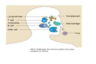
Complement • Three primary functions: • Lysis of antibody coated cells, such as bacteria and RBCs. • Mediation of opsonization, preparation of foreign cells for phagocytosis. • Generation of peptide fragments that regulate features of the inflammatory and immune response.
Importance in Blood Banking • Two major areas: • Some antigen-antibody complexes cause sufficient quantities of complement to be bound to RBCs to complete activation cycle, causing hemolysis. • Antigen-antibody complexes initiate complement binding in such a way that allows demonstration of the existence of such complexes by the use of serologic techniques