Download
1 / 56
590 likes | 618 Views
HLA system (MHC glycoproteins ). MHC glycoproteins class I (Major histocompatibility complex ).

E N D
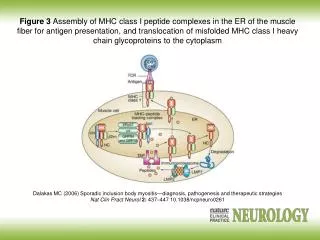
MHC glycoproteinsclass I (Major histocompatibilitycomplex) • MHCgpIpresentpeptide fragmentsfromitracellularproteins(which are produced by cell, includingviralpeptidesif are present) on the cell surfaceforcytotoxic T lymphocytes( CD8+) • Expressedon allnucleatedcells • 3 isotypesofclassical MHC gp. (HLA - A,-B,-C) • 3 isotypes non-classical MHC gp. (HLA - E,-F,-G; molecule CD1)
MHC gp I structure • MHC gp class I consists of transmembranechain a and associated b2microglobulin • a1, a2 - binding site for peptides • Peptide binding is necessary for a stable conformation of MHC gp
MHC gpI peptide binding • MHC gp I bindpeptideslong8 - 10 aminoacids • CertainMHC gpmoleculebindspeptidessharingidenticalstructuralfeaturesbindingmotif • Thebindingofendogenouspeptidesoccurs in theendoplasmicreticulumduringbiosynthesisof MHC gp I • These peptides are produced from intracellularproteins that are cleaved by theproteasomes
Non-classical MHC gp I • HLA - E,-F,-G; CD1 molecules • Structurally similar to classical MHC gp • Less polymorphic • Expressed only on some cells • They specialize in binding of specific ligands
HLA-EandHLA-G- expressed on the trophoblast cells • Complexes of HLA-E and HLA-G with peptides are recognized by NK cells inhibitory receptors and contribute to the tolerance of the fetus in utero
MHC glycoproteinsclass II • MHC gpII present peptide fragments from extracellular proteins on the cell surface for helper T lymphocytes (CD4+) • Expressed on the APC (dendritic cells, monocytes, macrophages, B lymphocytes) • 3 isotypes of MHC gpII (DR, DQ, DP)
MHC gp IIstructure • MHC gp II consist of 2 associated transmembrane chainsaand b • a1, b1 -binding site for peptide • Peptide binding is necessary for a stable coformation of MHC gp and ensure its long presentation on the cell surface
MHC gp II peptide binding • MHC gp II bindpeptideslong15 - 35 aminoacids • Certain MHC gpmoleculebindspeptidessharingidenticalstructuralfeatures–bindingmotif • Invariant chainblocksthebindingsiteforthepeptide • Exogenouspeptidesbinds to MHC gp II in theendosome • Peptide fragmentsfromendocytosedextracellularproteins
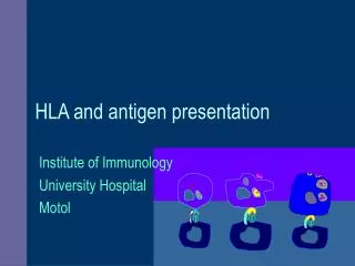
Antigen prezentation An antigen-presenting cell (APC) process foreign antigensand present them complexed withMHC‘s on their surfaces to T cells.
MHC glycoproteinspolymorphism • HLA complex is located on chromosome 6 • For MHC gp is typical high polymorphism (hundreds of different alelic forms of isotypes) • Codominant inheritance of alelic forms
MHC glycoproteinspolymorphism • Increases resistance to disease • Causes complications in the organ transplantation • Association of certain alleles with autoimmune diseases and increased susceptibility to infections
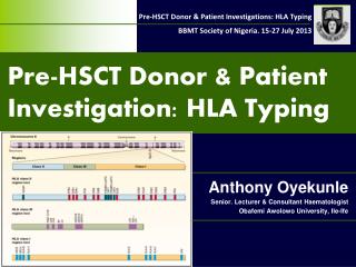
HLA typing = determminationof HLA antigens on thesurfaceoflymphocytes • Carry out during the testing before transplantation and in determination of paternity • serotyping • genotyping
Serotyping (microlymfocytotoxic test) • Allospecificserums(obtainedfrom multiple natal to 6 weeksafterbirth, orcommerciallypreparedsetsoftypingserums (monoclonalantibodies)) • Principle- theincubationoflymphocyteswithtypingserums in the presence ofrabbitcomplement, thenisaddedthevitaldyewhichstaineddeadcells - cellscarryingspecific HLA are killed by cytotoxic Ab againsttheAg, thepercentageofdeadcellsis a measureofserum toxicity (forcesandantileukocyteantibody titre) • Positive reactionisconsidered more than 10% deadcells(serologicaltypingcanbedonealso by flowcytometry)
Moleculargeneticmethods - genotyping a) PCR-SSP (Polymerasechainreactionwithsequentialspecificprimers) • Extracted DNA isused as a substrate in a set of PCR reactions • Each PCR reactioncontainsprimers pair specificfor a certainallele(orgroupofalleles) • Positive and negative reactions are evaluated by electrophoresis
Moleculargeneticmethods - genotyping b) PCR-SSO • PCR reactionwithsequence-specificoligonucleotides • Hybridizationwith enzyme orradiolabeledoligonucleotidesprobesspecificforindividualalleles
Molecular genetic methods - genotyping c) PCR-SBT • Sequencing based typing • We get the exact sequence of nucleotides, which compares with a database of known sequences of HLA alleles
T cells • Cellular component of antigen-specific mechanisms • Several subsets of T lymphocytes (TH1, TH2, Treg, TC…) • Regulation of immune processes and destruction of virus-infected cells or tumor cells • TCR recognize peptide-MHC complex • T cell are activated by APC
T cell development • T cells originate in bone marrow and then migrate to the thymus where they mature (abT lymphocytes), the final differentiation is after activation by antigen processed and presented by APC • gdT cells can develop outside the thymus (the minority population) • T cells are after activation stimulated to proliferation and differentiation into effector cells and memory cells
T cell development Pluripotenthematopoietic stem cells Pro-thymocytes– double negative T cells- are comingfromthe bone marrow to thethymus, wheretheybegin to rearrangeTCRbgenes, expressing on theirsurface, calledpre-TCR (Composedofbchain, pre-TCRaand CD3 complex), thenbeginTCRagenesrearrangement Corticalthymocytes – double positive T cells- express on theirsurfaceTCR (composedofchainsa, b and CD3) andCD4 and CD8 co-receptor (double positive T lymphocyte), atthisstageoccurstheselectionofautoreactivecellsandthecellswithdysfunctional TCR Medullarythymocytes (mature T cell) - retaintheexpressionofCD4 or CD8, thenmigrate to secondarylymphoidorgans
T cell selection • Negative selection- the elimination of autoreactive cells, when thymocytes binds strongly by their TCR complex of MHCgp with normal peptides (from autoantigens) which are presented on surface of thymic cells thymocyte receives signals leading to apoptotic cell death • Positive selection- the elimination of cells with dysfunctional TCR, positively are selected thymocytes that recognize MHC gp with low affinity, then maintain the expression of CD4 or CD8 (depending what class of MHC gp binds to the TCR). These mature T cells (Medullary thymocytes) leave the thymus and migrate to secondary lymphoid organs • 98% of pro-thymocytes in the thymus during its development dies
T cell surface markers • TCR - recognizes Ag peptide complexed with MHC gp • CD3 - TCR component, participation in signal transduction • CD4 or CD8 - co-receptors, binding to MHC gp • CD28 - costimulatory receptor, binds to CD80, CD86 on APC • CTLA-4 (CD152) - inhibitory receptor, binds to CD80, CD86
T cell subpopulations • ab-T lymphocytes- have TCRab, major type (95-98%), need thymus for development, recognize peptide antigens in the complex with MHC gp • gd-T lymphocytes- (2-5%) may develop outside the thymus, some are able to recognize native Ag, apply in defense of the skin and mucous membranes
ab T lymphocytes Expressing the CD4 coreceptor (co-receptor for MHC class II gp), precursors of helper T cells (TH),they can be classified according to the production of cytokines TH0 - produce a mixture of cytokines such as TH1 and TH2 TH1 - IL-2, IFNg (activates macrophages ) TH2 - IL-4, IL-5, IL-6, IL-10 (B lymphocytes assistance) TH3 – TGFb Treg - regulatory T cells arise in the thymus from a part of autoreactive lymphocytes, suppress the activity of autoreactive T cell clones (IL-10, TGFb)
ab T-lymphocytes Expressingthe CD8 co-receptor(co-receptor for MHC gpI), precursorsofcytotoxic T cells (TC) TC– recognizeanddestroycellsinfected by virusesorotherintracellularparasitesandsomecancercells
TCR • TCR (T cell receptor) isheterodimerconsistingofaandb (g,d) chains • associatedwithCD3 complex, whichisnecessaryforsignaltransduction • N-terminalpartsofaandb (g,d) chainsformthebindingsiteforAg
T cell activation • T cell are activated by APC (DC, monocyte, macrophage, B cell) • TCRrecognize peptide-MHC complex • TCR cooperate with coreceptors CD4 (binds to MHC gp II) or CD8 (binds to MHC gp I)
T cell activation • For full activation are necessary 2 signals • The first signal :TCR binding to peptide-MHC complex • The second signal comes from T cell co-stimulatory receptor CD28 which binds to CD80, CD86 on APC • Without costimulation, the T cell becomes anergic (prevention of inappropriate responses to self-peptides)
T cell activation Signal:TCR– MHC gp I(II)+Ag peptid (APC) Co-stimulating signal: CD 28 (T lymphocyte) – CD 80, CD 86 (APC)
TH1 immune response - inflammatoryreaction • TH1 cellscooperatewithmacrophagesandactivatethem(NO production - destroyintracellularparasites) • Activatedmacrophagessecretesomecytokines (IL-1, TNF, ...) that help to stimulate T cellsandstimulatelocalinflammation, whichhelpssuppressinfection • Interactionbetween TH1 cellsandmacrophagesis a fundamentalmechanismofdelayed-type immunopathologicalreactions (DTH Delayed-type hypersensitivity)
TH1 immune response • The infected macrophage produces protein fragments derived from intracellular parasites, some of which are presented on the surface in the complex with MHC gp class II • Macrophages and dendritic cells stimulated by certain microorganisms produce IL-12 • TH precursor, which detects the infected macrophage and receives signals via the TCR, CD 28 and receptor for IL-12 proliferates and differentiates into effector TH1 cells that produce IFNg and IL-2. • IFNg activates macrophage NO synthase IL-2 is growth factor for T cells
TH2 immune response – help to B cells • TH2 cells cooperate with B lymphocytes (which were stimulated by Ag) by cytokine production (IL-4, IL-5, IL-6, IL-10) and direct intercellular contact (CD 40L) • For stimulation of B lymphocytes is usually necessary cooperation between APC → TH2 cell → B lymphocyte • In minimal model, where the B cell becomes a good APC (CD80, CD86) is sufficient cooperation between TH2 cell → B lymphocyte
TH precursor, which detects the infected macrophage and receives signals through the TCR, CD 28 , IL-4 receptorand IL-2 receptor proliferates and differentiates in the effector TH2, which provide B lymphocytes auxiliary signals via secreted cytokines IL-4, IL-5, IL-6, IL-10 and molecule CD 40L, which bind to the costimulatory receptor on B lymphocytes CD 40 • Interaction between CD40 (B lymphocytes) and CD40L (TH2 cells) is essential for the initiation of somatic mutations, izotype switching and formation of memory cells • IL-4, IL-5, IL-6, IL-10: stimulation of B lymphocytes
Mutual regulation of activities TH1versus TH2 • Whether the TH precursor cell will develop into TH1 or TH2 decides cytokine ratio of IL-12 and IL-4 • IL-12 is produced by macrophages and dendritic cells stimulated by certain microorganisms • IL-4 is produced by activated basophils, mast cells and TH2 cells • TH1 cytokines (mainly IFNg) inhibit the development of TH2 and stimulate the development of TH1 (IL-2 stimulates also TH2) • Cytokines produced by TH2 (IL-4, IL-10) inhibit the development of TH1 and stimulate the development of TH2