Download
1 / 31
510 likes | 1.06k Views
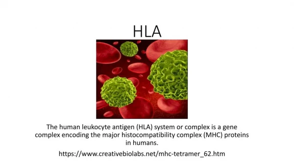
HLA TYPING. B Montague. HLA = Human Leukocyte Antigen system. HLA forms part of the Major Histocompatibiblity Complex (MHC) Found on the short arm of chromosome 6 MHC antigens are integral to the normal functioning of the immune response.

E N D
HLA TYPING B Montague
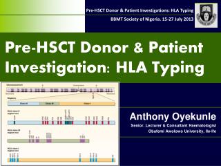
HLA = Human Leukocyte Antigen system • HLA forms part of the Major Histocompatibiblity Complex (MHC) • Found on the short arm of chromosome 6 • MHC antigens are integral to the normal functioning of the immune response. • Essential role of HLA antigens lies in the control of self recognition and thus defence against micro-organisms and surveillance.
Interesting facts • HLA comprises two classes: Class I Class II . Class I A,B,C most significant (other loci eg E,F,G,H etc are not so important in transplantation) . Expressed on most nucleated cells . Have soluble form in plasma . Are adsorbed onto platelets (some antigens more readily than others)
Erythrocytes will adsorb some Class I antigens viz. Bg blood group system (B7,A28, B57….) HLA B most polymorphic system and studies have shown is most significant followed by A and then C 45Kd glycoprotein comprising three heavy chain domains, non-covalently associated More interesting facts
with beta-2-microglobulin (coded chromosome 15) which plays an important role in the structural support of the heavy chains. • Class I molecules are assembled within the cell and ultimately sit on the cell surface with a section inserted into the lipid bilayer of the cell membrane and a short cytoplasmic tail where they present antigen in the form of peptide to cytotoxic T (CD8+) cells
HLA Class II five loci DR, DQ, DP, DM and DO HLA DR, DQ, DP most significant Expressed on B lymphocytes, activated T lymphocytes, macrophages, endothelial cells ie immune competent cells. Comprise 2 chains encoded by HLA genes, alpha and beta each with 2 domains. Hypervariable region is in the beta 1 domain
HLA Class II present peptide in the cleft to helper T (CD4+) cells. Thus Class II presentation involves the helper-function of setting up a general immune reaction involving cytokine, cellular and humoral defence. • The role of Class II in initiating a general immune response is why they only need to be present on immunologically active cells.
TYPING METHODS • SEROLOGY used to be the ‘gold’ standard. Now being superceded by molecular techniques as they become more robust and time efficient • CELLULAR rarely used now. Orginally used for Class II typing • MOLECULAR fast becoming the method of choice. Many laboratories test of choice.
SEROLOGY • Complement Dependent Cytotoxicity (CDC) • Viable peripheral blood lymphocytes are obtained by discontinous density gradient centrifugation using Ficoll / Tryosil or Ficoll / Sodium Metrizoate at a density of 1.077 at 19º - 22ºC. • Microlymphocytotoxic test: 3 stages
Microlymphocyototoxic test • 1.Viable lymphocytes are incubated with HLA specific antibodies. If the specific antigen is present on the cell the antibody is bound. • 2.Rabbit serum as a source of complement is added, incubate. If antibody is bound to the HLA antigen on the cell surface it activates the complement which damages the cell membrane making it permeable to vital stains.
Microlymphocyototoxic test 2 • 3.Results are visualised by adding dye usually a fluorochrome eg Ethidium Bromide although both Trypan Blue and Eosin have been used in the past. • If the reaction has taken place the EB enters the cell and binds to the DNA. • For ease double staining is normally used. We use a cocktail of Ethidium Bromide and Acridine Orange, quenched using Bovine Haemoglobin to allow simultaneous visualisation of both living and dead cells.
Microlymphocytotoxicity test 3 • Test is left for 10 minutes and then read using an inverted fluorescient microscope. • A mixture of T and B lymphocytes can be used for HLA Class I typing. • B lymphocytes are required for HLA Class II typing by serology. (Normal population 85-90% T and 10-15% B cells) • This can be achieved using a number of methods.
Microlymphocytotoxicity test 4 • In the past neuraminidase treated sheep red blood cell rosetting and nylon wool have been used. • Immunomagnetic bead separation is the current method of choice. • It utilises polystyrene microspheres with a magnetisable core coated in monoclonal antibody for a HLA Class II b chain monomorphic epitope. Positive selection.
Pros and cons • Pros: • Easily performed does not require expensive equipment. • Takes around three hours to perform • Low level resolution, with good antisera reliable results • Cons: • Requires large volumes of blood • Requires viable lymphocytes • Difficult to find good antisera for rarer antigens in different populations
Cellular typing • Not / Rarely used by laboratories these days. • Requires panels of homozygous typing cells. • Cell culture method therefore takes a long time. Labour intensive involves use of radioisotopes.
Molecular • All commonly used molecular methods require good quality genomic DNA. There are numerous methods for extraction of DNA from whole blood. • There are ‘in house’ methods based on Miller et al’s Salting Out which are cheap and easy but labour intensive. • There are also numerous commercial kits available such as individual matrix capture columns, beads and semi automated systems. This however can increase the cost per extraction from around 65p to £3.60p.
Molecular 2 • All methods rely on DNA extraction from the nucleated cells following cell lysis and protein digestion.
Molecular Methods • The application of molecular techniques to HLA typing began around 1987 when the Southern Blot technique was used to identify restriction fragment length polymorphisms (RFLP’s) associated with known serological DR/DQ and cellular Dw defined specifities. • Around 1992 polymerase chain reaction (PCR) methods were developed. • Most methods currently used have a PCR element within the technique.
Molecular Methods 2 • PCR • Three steps per cycle– denaturation, annealing and extension. Amplification is exponential yielding 2 power n where n = number of cycles. • The introduction of the programmable Thermal Cycler revolutionised the use of PCR within the routine laboratory.
Molecular Methods 3 • PCT SSP (Sequence Specific Priming) • Can be used for HLA Class I and II typing using a panel of primer pairs either for low to medium resolution whereby primers amplify groups of alleles or high resolution whereby primer pairs amplify specific alleles. Each PCR reaction takes place in a separate tube therefore the number of tubes depends on the level of resolution. Each tube also contains a pair of primers for part of the human growth hormone gene as an internal control. These are at a much lower concentration thus do not compete with specific primers.
Molecular Methods 4 • Electrophoresis is used following amplification. PCR product is run out on an agarose gel containing ethidium bromide. Each product moves according to its size and is compared to a molecular weight marker. • Interpretation: every tube should produce an identical sized product as internal control and either a specific band or not dependent on whether the allele(s) is/are present or not. • Results are visualised using 312nm UV transillumination and recorded either by video imaging or polaroid photograghy.
Molecular methods 5 • PCR SSOP ( Sequence Specific Oligonucleotide Probes) • ‘Dot blot’ in house method usually whereby one labels ones own probes with Digoxigenin • ‘Reverse dot blot’ normally commercial where specific oligonucleotide probes are attached to a nylon membrane. Dynal and Innotrans for example produce such kits.
Molecular Methods 6 • Amplification: DNA of interest is amplified by a single pair of biotinylated primers which flank the whole of exon eg exon 2 of the HLA DRB1 gene. PCR amplifies all the alleles in the exon. • Hybridisation: PCR product is denatured and then added to a ‘well’ containing the nylon membrane with the bound probes and incubated with hybridisation buffer . PCR product hybridises to probes with complementary sequences. • Excess product is washed away during a series of wash steps. • Temperature is VERY important during these stages. • Visualisation of results is achieved by incubating with a conjugate and enzyme often streptavidin and horse radish peroxidase which binds to the biotin of the PCR product and then adding a substrate. Band with PCR product turn blue. • Strips will have internal control bands to show the test has worked. • Interpretation is usually achieved by entering the band pattern into a computer programme. • This is an excellent method for low resolution batch testing. • Can be semi automated.
Pros and Cons • Pros: • Does not require viable cells • Samples do not have to arrive in the lab the day they are taken • PCR SSOP good for batch testing • Can be semi automated • Cons • Requires good quality DNA • Require a degree of redundancy within the primers used • Sequence of alleles must be known.
Molecular Methods 7 • Sequence Based Typing (SBT) • DNA sequencing is the determination of the sequence of a gene and thus is the highest resolution possible. Sequence based typing involves PCR amplification of the gene of interest eg HLA DRB1 followed by determination of the base sequence. The sequence is then compared with a database of DRB1 gene sequences to find comparable sequences and assign alleles. This method also allows for detection on new alleles.
Molecular Methods 8 • Other molecular methods: • Reference Strand Conformational Analysis (RSCA) Offers sequence level typing without the need to sequence. Assigns HLA type on the basis of accurate measurement of conformation ie. shape dependent on DNA mobility in polyacrylamide gel electrophoresis (PAGE). Complex and difficult technique not taken up by labs for routine use.
Molecular Methods 9 • Luminex technology – SSOP based. Just beginning to be introduced into laboratories for routine use on non urgent samples.
Nomenclature • In the 1950’s Dausset, Rose and van Rood amongst others described leukoagglutinating antibodies in the serum of patients who had been pregnant or transfused and that patterns of reaction were observed against random cell panels. • Numbers of investigators increased and it was realised that collaboration was needed to develop a standardised nomenclature and typing methods. • Out of this desire was born the first International Workshop in Durham, North Carolina in 1964. • Since then there have been 14 such workshops.
Nomenclature 2 • These workshops led to a logical and consistent if somewhat complex nomenclature. • Within HLA A and B loci you will not find the same number used to describe an antigen eg HLA-A1 there is NOT an HLA-B1 or HLA-B8 there is not an HLA-A8 and so on. • In serology HLA C retains ‘w’ to differentiate from the complement cascade
Nomenclature 3 • HLA-A identifies HLA A locus • HLA-A1 serologically defined antigen • HLA-A* asterisk denotes HLA alleles defined by molecular methods • HLA-A*01 2 digit resolution denotes a group of alleles corresponds usually to serological group – low resolution • HLA-A*0101 4 digit resolution – sequence variation between alleles results in amino acid substitutions • HLA-A010101 6 digit resolution – non coding variation: sequence changes synonymous no amino acid substitution
Nomenclature 4 • HLA-A01010101 8 digit resolution – sequence variation occurs within the introns or 5’ / 3’ extremities of the gene • HLA-A01010102N Null allele • Alphabetical suffix • N Null allele • L Low level expression • A Aberrant expression • C Molecule present in cytoplasm only • S Secreted molecule present only as soluble form