Download
1 / 29
290 likes | 545 Views
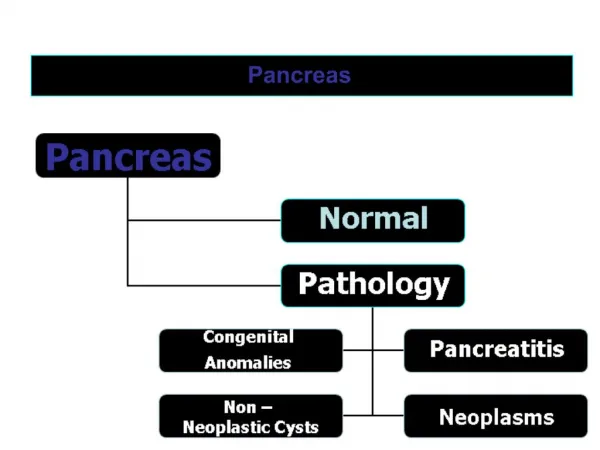
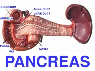
Normal pancreas. Pathology of pancreas. P ancreatitis . Inflammation of the pancreas May be diffuse or focal Acute or chronic

E N D
Pancreatitis Inflammation of the pancreas May be diffuse or focal Acute or chronic Occur due to response to the destruction of pancreatic tissue by its own digestive enzymes ( autodigestion ) which have been released from damaged pancreatic cells .
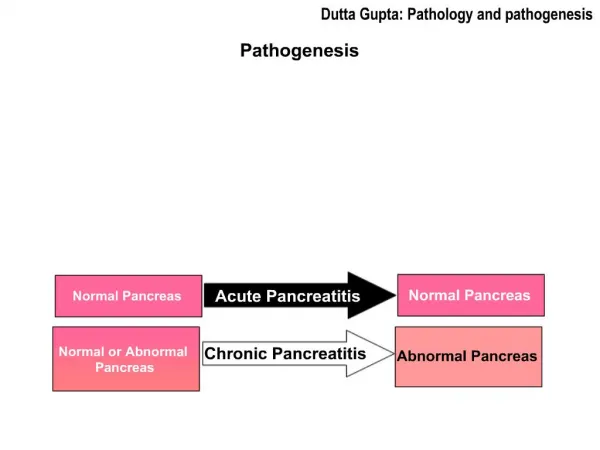
Acute pancreatitis Acute inflammation causes the pancreatic tissue necrosed , releasing the pancreatic enzymes which destroy the pancreatic tissue and the capillary walls and entering the blood stream. Causes :- 1- Biliary calculi , it is the most common cause 2- Alcoholism 3- Trauma 4- Drug induced (rare cause) 5- Infection e.g mumps ( rare cause )
Acute pancreatitis Clinical picture :- Sever epigastric pain Abdominal distension Nausea or vomiting In mild cases the patient may recover spontaneously Untreated cases may lead to peritonitis Raised levels of amylase and lipase
Acute pancreatitis What is the role of ultrasound :_ The main role of U.S in cases of acute pancreatitis try to detect the cause of pancreatitis .
In mild cases there are no changes by ultrasound. Ultrasound appearance:- • In sever cases • Hypoechoic enlarged pancreas • CBD dilatation if there is enlargment of pancreatic head • A collection or pseudocystdue to leakage out of digestive pancreatic enzymes • Pseudocyst May be seen in the lesser sac near the tail of pancreas , within the pancreatic tissue itself , anywhere in the abdomen .
Acute pancreatitis Pseudocyst may be echofree but may contain echoes from tissue debris and may be loculated . Pancreatic abscess may be occur due to infection
Acute pancreatitis Enlarged edematous pancreas “P” 4cm pseudocyst “C”
Acute pancreatitis The pancreas (P) is draped over the splenic vein (SV), which is hypoechoic and swollen, with a rim of fluid around its edge (white arrows). A is the aorta and IVC the inferior vena cava
Chronic pancreatitis Causes :- May be occur on top of acute pancreatitis Alcohol abuse The normal pancreatic tissue is replaced by fibrosis .
Chronic pancreatitis Clinical picture :- Pain Steatorrhoea Serum enzyme levels are less elevated than in acute disease.
Chronic pancreatitis Ultrasound appearance :- Abnormally hyperechogenicity of pancreas Atrophied and lobulation of pancreas Dilatation of the pancreatic duct Pancreatic calcification
Chronic pancreatitis Chronic Pancreatitis, echogenic irregular appearance to the body of the pancreas
Pancreatic carcinoma Pancreatic carcinoma is a major cause of cancer related death Clinical picture Presenting symptoms depends on the size of the lesion ,and its position within the pancreas Most pancreatic tumor ( 60% ) are found in the head of the pancreas and the patient present by obstructive jaundice ……Why ? Carcinoma which located in the body and tail of pancreas do not cause obstructive jaundice .
Pancreatic carcinoma Ultrasound appearance :- Solid hypoechoic or mixed echogenicity with an irregular border Cystic carcinoma appear as multiseptate cystic masses with associated solid components
Pancreatic cysts The pancreatic cysts may be Benign pancreatic cysts Cystic carcinoma ( rare ) Pseudocyst associated with acute pancreatitis .
Benign pancreatic cysts Rare and tend to associated with other conditions as polycystic disease . Single or multiple Echo free small cavities filled with fluid
Trauma of pancreas Pancreas is exposed to trauma especially in road traffic accidents Ultasound is senstive to detect leakage of pancreatic juice into the abdominal cavity (caused by rupture of pancreatic duct) and hematoma caused by laceration of the pancreas
Calcification in the pancreas Calcification within the pancreas can produce acoustic shadowing but if it is very small there may only be bright descrete echoes without shadowing Causes of pancreatic calcifications Chronic pancreatitis Calculi in the pancreatic dust Biliary calculi in the distal common bile duct can be mistaken for pancreatic calcification , there is usually dilatation of the proximal bile duct .
Dilatation of pancreatic duct The maximal internal diameter of pancreatic duct is 2 mm Causes of dilatation of pancreatic duct:- Tumor of head of the pancreas Calculus in the CBD Calculus in the pancreatic duct Chronic pancreatitis
Diffuse enlargement of the pancreas Causes :- Acute pancreatitis Acute on top of chronic pancreatitis
Focal enlargement of pancreas Causes :- Focal pancreatitis Pancreatic tumor How can you differentiate Serum amylaze is elevated with pancreatitis In some cases biopsy is needed to differentiate • Previous history of pancreatitis • Normal tumor marker level
Small pancreas Old age Chronic pancreatitis