Download
1 / 37
430 likes | 753 Views
Pathology of rheumatic fever, IE & Valvular diseases DR. AMMAR AL-RIKABI / Dr Shaesta Naseem. Rheumatic fever (RF). Rheumatic fever (RF). Acute, immunologically mediated, multisystem inflammatory disease. Involves heart, blood vessels, joints, subcutaneous tissue and CNS .

E N D
Pathology of rheumatic fever, IE & Valvular diseasesDR. AMMAR AL-RIKABI /DrShaestaNaseem
Rheumatic fever (RF) • Acute, immunologically mediated, multisystem inflammatory disease. • Involves heart, blood vessels, joints, subcutaneous tissue and CNS . • Occurs in 3% of patients, a few weeks after an episode of group A streptococcal pharyngitis. • Most often in children between ages 5 and 15. • Deforming fibrotic valvular abnormalities (esp MS) are important cardiac complications.
Pathologic sequence and key morphologic features of acute RHD
Clinical features • RF is characterized by a constellation of findings that includes as major manifestations: • (1) migratory polyarthritis of the large joints, • (2) pancarditis, • (3) subcutaneous nodules, • (4) erythema marginatum of the skin, and • (5) Sydenham chorea, a neurologic disorder with involuntary rapid, purposeless movements. • The diagnosis is established by Jones criteria
PATHOLOGY of RF • Pathological hallmark : Aschoff bodies. • Aschoff bodies consist of foci of fibrinoid degeneration surrounded by lymphocytes (primarily T cells), occasional plasma cells, and plump activated macrophages called Anitschkow cells. • Anitschkow cells : have abundant cytoplasm and central round-to-ovoid nuclei in which the chromatin is disposed in a central, slender, wavy ribbon ("caterpillar cells") • It may become multinucleated.
Aschoff nodule and Anitschkow cell • During acute RF, diffuse inflammation and Aschoff bodies may be found in any of the three layers of the heart, causing pericarditis, myocarditis, or endocarditis (pancarditis)
. Rheumatic Heart Disease
Rheumatic endocarditis • Inflammation results in fibrinoid necrosis within the cusps or along the tendinous cords. • Overlying these necrotic foci are small (1- to 2-mm) vegetations, called verrucae, along the lines of closure. • Subendocardial lesions, exacerbated by regurgitant jets, may induce irregular thickenings called MacCallum plaques, usually in the left atrium.
In chronic disease the mitral valve is virtually always involved. • Mitral valve in chronic RHD are leaflet thickening, commissural fusion and shortening, and thickening and fusion of the tendinous cords
Small vegetations (verrucae) are visible along the line of closure of the mitral valve leaflet Aschoff body in myocardium Mitral stenosis with diffuse fibrous thickening and distortion of the valve leaflets and commissural fusion (arrows, C), and thickening of the chordae tendineae Rheumatic aortic stenosis
. Schematic representation of the anatomic regions of involvement and location of vegetations in rheumatic endocarditis.
. Rheumatic Heart Disease
Infective endocarditis (IE) • It is a serious infection characterized by colonization or invasion of the heart valves or the mural endocardium by a microbe. • This leads to the formation of vegetations • Most cases are caused by bacterial infections (bacterial endocarditis).
Clinical presentation and complications • Acute: • Fever, rigor, malaise • Large vegetation => emboli: • Infarction • Metastatic infection • Distant organs like spleen, brain or heart • Kidney: Ag-Ab complex -> GN-> nephrotic syndrome or Renal failure • Congestive heart failure due to valve disease • Can lead to ring abscess and perforation of the aorta and myocardium • Death up to 60% • Subacute: • Insidious • Splenomegaly • Non specific : fever, weight loss
The hallmark of IE is the presence of friable, bulky, potentially destructive vegetations . • The aortic and mitral valves are the most common sites of infection. • Vegetations containing fibrin, inflammatory cells, and bacteria on the heart valves • Vegetation sometimes erode into the underlying myocardium and produce an abscess (ring abscess).
. Infective endocarditis
Infective (bacterial) endocarditis. Acute endocarditis of congenitally bicuspid aortic valve Endocarditis of mitral valve Extensive acute inflammatory cells and fibrin. Healed endocarditis
NONINFECTED VEGETATIONS(sterile) • Nonbacterial thrombotic endocarditis and the endocarditis of SLE, called Libman-Sacks endocarditis. • NBTE is often encountered in debilitated patients, such as those with cancer or sepsis. • It frequently occurs concomitantly with deep venous thromboses, pulmonary emboli.
NBTE • NBTE is characterized by the deposition of small sterile thrombi on the leaflets of the cardiac valves. • The lesions are 1 mm to 5 mm in size, and occur singly or multiply along the line of closure of the leaflets or cusps. • Histologically they are composed of bland thrombi that are loosely attached to the underlying valve. • The vegetation are not invasive and do not elicit any inflammatory reaction.
Nonbacterial thrombotic endocarditis (NBTE). A, Nearly complete row of thrombotic vegetations along the line of closure of the mitral valve leaflets (arrows).B, Photomicrograph of NBTE, showing bland thrombus, with virtually no inflammation in the valve cusp (c) or the thrombotic deposit (t). The thrombus is only loosely attached to the cusp (arrow
Comparison of the four major forms of vegetative endocarditis. The rheumatic fever phase of rheumatic heart disease (RHD) is marked by small, warty vegetations along the lines of closure of the valve leaflets. Infective endocarditis (IE) is characterized by large, irregular masses on the valve cusps that can extend onto the chordae . Nonbacterial thrombotic endocarditis (NBTE) typically exhibits small, bland vegetations, usually attached at the line of closure. One or many may be present . Libman-Sacks endocarditis (LSE) has small or medium-sized vegetations on either or both sides of the valve leaflets.
. Cardiac squeal of infective endocarditis Extra-cardiac Complications
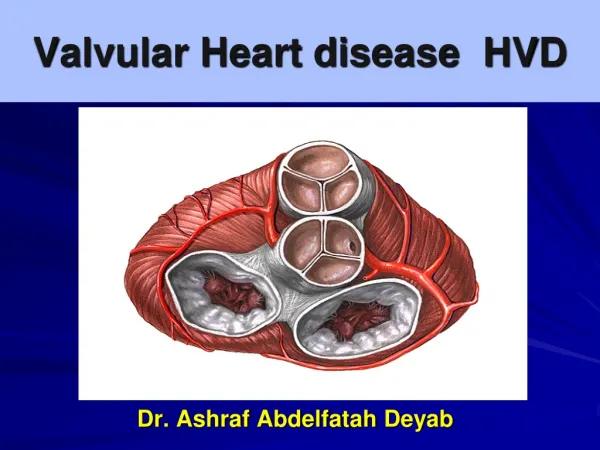
Valvular Heart Disease • Can come to clinical attention due to stenosis, insufficiency (regurgitation or incompetence),or both. • Stenosis is the failure of a valve to open completely, which impedes forward flow. • Insufficiency, in contrast, results from failure of a valve to close completely, thereby allowing reversed flow.
The most frequent causes of the major functional valvular lesions are: • •Aortic stenosis: calcification of anatomically normal and congenitally bicuspid aortic valves • •Aortic insufficiency: dilation of the ascending aorta, usually related to hypertension and aging • •Mitral stenosis: rheumatic heart disease • •Mitral insufficiency: myxomatous degeneration (mitral valve prolapse)
Calcific Aortic Stenosis • The most common of all valvular abnormalities • The consequence of age-associated "wear and tear. • heaped-up calcified masses within the aortic cusps . • It ultimately protrude preventing the opening of the cusps. • Microscopically, the layered architecture of the valve is largely preserved.
Aortic Stenosis • Valve becomes stiff and fibrotic, impeding blood flow with LV contraction • Results in LV hypertrophy, increased O2 demands, and pulmonary congestion. • Causes – rheumatic fever, congenital, arthrosclerosis Atherosclerosis and calcification is primary cause in the elderly
Aortic Stenosis Symptoms • Angina • Syncope • Congestive Heart Failure (CHF) • Complications – right sided heart failure, pulmonary edema, and A-fib
Aortic Regurgitation Etiologies • Abnormalities of the Leaflets • Rheumatic, Bicuspid, Degenerative • Endocarditis • Dilation of the Aortic Annulus • Aortic Aneurysm / Dissection • Inflammatory • Inheritable (Marfans, Osteogensis Imperfecta)
Mitral Stenosis Etiologies • Rheumatic – almost all cases in adults • Mitral Annular Ca+ - massive (rare) • Congenital – rare
Mitral Regurgitation Symptoms • Fatigue and weakness • Dyspnea and orthopnea • Right sided HF