Download
1 / 88
880 likes | 890 Views
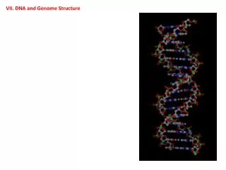
VII. DNA and Genome Structure. VII. DNA and Genome Structure A. Search for the Genetic Information. VII. DNA and Genome Structure A. Search for the Genetic Information 1. Early Work

E N D
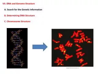
VII. DNA and Genome Structure A. Search for the Genetic Information
VII. DNA and Genome Structure A. Search for the Genetic Information 1. Early Work a. Miescher – 1868 – isolated nuclein from the nucleus of cells. An acidic, nitrogen rich material.
VII. DNA and Genome Structure A. Search for the Genetic Information 1. Early Work a. Miescher – 1868 – isolated nuclein from the nucleus of cells. An acidic, nitrogen rich material. b. Levene - 1910 – Chromosomes consist of DNA and proteins. DNA was very simple (4 nucleotides) whereas proteins were very complex (21 amino acids).
VII. DNA and Genome Structure A. Search for the Genetic Information 1. Early Work a. Miescher – 1868 – isolated nuclein from the nucleus of cells. An acidic, nitrogen rich material. b. Levene - 1910 – Chromosomes consist of DNA and proteins. DNA was very simple (4 nucleotides) whereas proteins were very complex (21 amino acids). Levene found that these nucleotides were in approximately an even ratio, and he hypothesized a very simple “tetranucleotide” structure that was similar over it’s length.
VII. DNA and Genome Structure A. Search for the Genetic Information 1. Early Work a. Miescher – 1868 – isolated nuclein from the nucleus of cells. An acidic, nitrogen rich material. b. Levene - 1910 – Chromosomes consist of DNA and proteins. DNA was very simple (4 nucleotides) whereas proteins were very complex (21 amino acids). Levene found that these nucleotides were in approximately an even ratio, and he hypothesized a very simple “tetranucleotide” structure that was similar over it’s length. Given that the genetic system must encode the diversity of life, it seemed likely that the more complex molecule (proteins) was responsible.
VII. DNA and Genome Structure A. Search for the Genetic Information 1. Early Work a. Miescher – 1868 – isolated nuclein from the nucleus of cells. An acidic, nitrogen rich material. b. Levene - 1910 – Chromosomes consist of DNA and proteins. DNA was very simple (4 nucleotides) whereas proteins were very complex (21 amino acids). Levene found that these nucleotides were in approximately an even ratio, and he hypothesized a very simple “tetranucleotide” structure that was similar over it’s length. Given that the genetic system must encode the diversity of life, it seemed likely that the more complex molecule (proteins) was responsible. c. Chargaff – 1940’s – [A] = [T], [C] = [G]; disproving Levene’s model.
VII. DNA and Genome Structure A. Search for the Genetic Information 1. Early Work 2. Major Experiments
VII. DNA and Genome Structure A. Search for the Genetic Information 1. Early Work 2. Major Experiments a. Griffiths – 1927 Streptococcus pneumoniae causes pneumonia, meningitis, sepsis Virulent strain has a polysaccharide capsule that protects the cell from being engulfed by white blood cells… and it makes them appear smooth (IIIS).
VII. DNA and Genome Structure A. Search for the Genetic Information 1. Early Work 2. Major Experiments a. Griffiths – 1927 Streptococcus pneumoniae causes pneumonia, meningitis, sepsis Non-virulent strain has no capsule and are killed by the immune system; they are ‘rough’ (IIR).
VII. DNA and Genome Structure A. Search for the Genetic Information 1. Early Work 2. Major Experiments a. Griffiths – 1927 Streptococcus pneumoniae causes pneumonia, meningitis, sepsis If virulent IIIS are killed by heat, they can be injected without causing disease.
VII. DNA and Genome Structure A. Search for the Genetic Information 1. Early Work 2. Major Experiments a. Griffiths – 1927 Streptococcus pneumoniae causes pneumonia, meningitis, sepsis If virulent IIIS are killed by heat, they can be injected without causing disease. Griffith found that a combination of LIVE IIR and DEAD IIIS, both non-virulent independently, would kill the mouse.
VII. DNA and Genome Structure A. Search for the Genetic Information 1. Early Work 2. Major Experiments a. Griffiths – 1927 Streptococcus pneumoniae causes pneumonia, meningitis, sepsis If virulent IIIS are killed by heat, they can be injected without causing disease. Griffith found that a combination of LIVE IIR and DEAD IIIS, both non-virulent independently, would kill the mouse. Concluded that the IIR received a HERITABLE ‘transforming factor’ from dead IIIS cells, and turned into live IIIS cells.
VII. DNA and Genome Structure A. Search for the Genetic Information 1. Early Work 2. Major Experiments a. Griffiths – 1927 Streptococcus pneumoniae causes pneumonia, meningitis, sepsis Thought it was a chemical that induced capsule formation.
VII. DNA and Genome Structure A. Search for the Genetic Information 1. Early Work 2. Major Experiments a. Griffiths – 1927 b. Dawson – 1931 Transformation in vitro (test tube)
VII. DNA and Genome Structure A. Search for the Genetic Information 1. Early Work 2. Major Experiments a. Griffiths – 1927 b. Dawson – 1931 Transformation in vitro (test tube) c. Alloway – 1933 Transformation with an extract from hk-IIIS – don’t even need the intact cells to cause a HERITABLE change in the live IIRIIIS What causes this heritable change: DNA, RNA, or protein?
2. Major Experiments d. Avery, McCarty, and MacLeod - 1944 • Took hk-IIIS extract and added live IIR – got transformation (control).
2. Major Experiments d. Avery, McCarty, and MacLeod - 1944 • Took hk-IIIS extract and added live IIR – got transformation (control). • Took hk-IIIS and added proteases that destroy proteins – got transformation; Transforming factor is NOT a PROTEIN
2. Major Experiments d. Avery, McCarty, and MacLeod - 1944 • Took hk-IIIS extract and added live IIR – got transformation (control). • Took hk-IIIS and added proteases that destroy proteins – got transformation; Transforming factor is NOT a PROTEIN • Took this solution, added RNAases – got transformation; Transforming factor is NOT an RNA
2. Major Experiments d. Avery, McCarty, and MacLeod - 1944 • Took hk-IIIS extract and added live IIR – got transformation (control). • Took hk-IIIS and added proteases that destroy proteins – got transformation; Transforming factor is NOT a PROTEIN • Took this solution, added RNAases – got transformation; Transforming factor is NOT an RNA • Added DNAases – NO TRANSFORMATION; transforming factor is DNA.
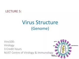
2. Major Experiments d. Hershey and Chase - 1952 • Viruses replicate within a bacterium… requiring the replication of the genetic information.
2. Major Experiments d. Hershey and Chase - 1952 • Viruses replicate within a bacterium… requiring the replication of the genetic information. • Viruses are about 50% DNA and 50% protein. Which goes inside the cell to cause change?
2. Major Experiments d. Hershey and Chase - 1952 • Viruses replicate within a bacterium… requiring the replication of the genetic information. • Viruses are about 50% DNA and 50% protein. Which goes inside the cell? • Labelled proteins with radioactive sulfur and DNA with radioactive phosphorus by growing virus on labelled bacteria for one generation.
2. Major Experiments d. Hershey and Chase - 1952 4) Then, they exposed normal bacteria to these differentially labelled viruses.
2. Major Experiments d. Hershey and Chase - 1952 4) Then, they exposed normal bacteria to these differentially labelled viruses. 5) Then they shook the solutions, separating the viral component from the bacterial component.
2. Major Experiments d. Hershey and Chase - 1952 4) Then, they exposed normal bacteria to these differentially labelled viruses. • Then they shook the solutions, separating the viral component from the bacterial component. • Both replicates confirmed that only DNA, and not protein, entered the cell and must be responsible for orchestrating viral reproduction. DNA is the genetic information.
VII. DNA and Genome Structure A. Search for the Genetic Information 1. Early Work 2. Major Experiments 3. Other Evidence
VII. DNA and Genome Structure A. Search for the Genetic Information 1. Early Work 2. Major Experiments 3. Other Evidence a. Mutagenesis The wavelengths of radiation that cause damage to the genetic information are the wavelengths absorbed by DNA, not proteins.
VII. DNA and Genome Structure A. Search for the Genetic Information 1. Early Work 2. Major Experiments 3. Other Evidence a. Mutagenesis b. Recombinant DNA Technology 1986 – gene for luciferase (from fireflies) was transferred to plant embryos. When they grew, and then were injected with luciferin (the enzymes substrate), the action of the enzyme (oxidation of luciferin) releases light.
VII. DNA and Genome Structure A. Search for the Genetic Information 1. Early Work 2. Major Experiments 3. Other Evidence a. Mutagenesis b. Recombinant DNA Technology c. RNA is the genetic information in some viruses RNA injected by virus can act directly (TMV), or can be copied into DNA (retroviruses) and inserted into the hosts genome and inherited during host cell replication (HIV).
VII. DNA and Genome Structure A. Search for the Genetic Information B. Determining DNA Structure
VII. DNA and Genome Structure A. Search for the Genetic Information B. Determining DNA Structure 1. Background Work: - Chargaff’s ratios
VII. DNA and Genome Structure A. Search for the Genetic Information B. Determining DNA Structure 1. Background Work: - Chargaff’s ratios - Astbury’s 3.4A periodicity
VII. DNA and Genome Structure A. Search for the Genetic Information B. Determining DNA Structure 1. Background Work: 2. Race for the Prize: a. Linus Pauling (CalTech) – Nobelist for describing helical structure of proteins, turned his attention to DNA.
VII. DNA and Genome Structure A. Search for the Genetic Information B. Determining DNA Structure 1. Background Work: 2. Race for the Prize: a. Linus Pauling (CalTech) – Nobelist for describing helical structure of proteins, turned his attention to DNA. He used X-Ray crystallography, and with impure samples of DNA, suggested DNA was a triple-helix…
VII. DNA and Genome Structure A. Search for the Genetic Information B. Determining DNA Structure 1. Background Work: 2. Race for the Prize: a. Linus Pauling b. Maurice Wilkins and Rosalind Franklin - The King College Lab, Univ. of London. - They had a more purified sample of DNA, but lab tensions made their supervisor assign Wilkins the ‘B’ form and Franklin the ‘A’ form. Wilkins concluded that the B form was helical; Franklin did not agree.
VII. DNA and Genome Structure A. Search for the Genetic Information B. Determining DNA Structure 1. Background Work: 2. Race for the Prize: a. Linus Pauling b. Maurice Wilkins and Rosalind Franklin - However, her subsequent work and beautiful x-rays ultimately convinced her of a double-helical structure… submitted to journals in March 1953 but without describing a specific model. Critical contributions were confirming Astbury’s 3.4A periodicity, and finding a larger periodicity at 34.0A.
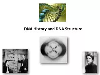
VII. DNA and Genome Structure A. Search for the Genetic Information B. Determining DNA Structure 1. Background Work: 2. Race for the Prize: a. Linus Pauling b. Maurice Wilkins and Rosalind Franklin c. Francis Crick and James Watson - Cavendish Lab, Cambridge University.
VII. DNA and Genome Structure A. Search for the Genetic Information B. Determining DNA Structure 1. Background Work: 2. Race for the Prize: a. Linus Pauling b. Maurice Wilkins and Rosalind Franklin c. Francis Crick and James Watson - Cavendish Lab, Cambridge University. - Crick was the crystallographer and a modeller. - Were working on helical structures with the ‘backbone’ on the inside. On seeing Franklin’s picture 51 in January 1953, they changed direction and ultimately produced a model of DNA that explained Franklin’s regularities and Chargaff’s Ratios.
VII. DNA and Genome Structure A. Search for the Genetic Information B. Determining DNA Structure 1. Background Work: 2. Race for the Prize: a. Linus Pauling b. Maurice Wilkins and Rosalind Franklin c. Francis Crick and James Watson - Cavendish Lab, Cambridge University. - Crick was the crystallographer and a modeller. - Were working on helical structures with the ‘backbone’ on the inside. On seeing Franklin’s picture 51 in January 1953, they changed direction and ultimately produced a model of DNA that explained Franklin’s regularities and Chargaff’s Ratios. d. 1958 – Franklin dies of ovarian cancer; probably related to her x-ray work.
VII. DNA and Genome Structure A. Search for the Genetic Information B. Determining DNA Structure 1. Background Work: 2. Race for the Prize: a. Linus Pauling b. Maurice Wilkins and Rosalind Franklin c. Francis Crick and James Watson - Cavendish Lab, Cambridge University. - Crick was the crystallographer and a modeller. - Were working on helical structures with the ‘backbone’ on the inside. On seeing Franklin’s picture 51 in January 1953, they changed direction and ultimately produced a model of DNA that explained Franklin’s regularities and Chargaff’s Ratios. d. 1958 – Franklin dies of ovarian cancer; probably related to her x-ray work. e. 1962 – Nobel Prizes for Crick, Watson, and Wilkins
VII. DNA and Genome Structure A. Search for the Genetic Information B. Determining DNA Structure 1. Background Work: 2. Race for the Prize: 3. The Structure of DNA
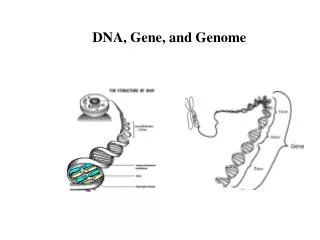
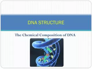
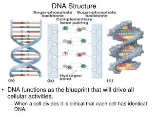
3. The Structure of DNA (and RNA) - basic unit is a “nucleotide”, that has three parts: i. pentose sugar:
3. The Structure of DNA (and RNA) - basic unit is a “nucleotide”, that has three parts: i. pentose sugar: ii. Nitrogenous base:
3. The Structure of DNA (and RNA) - basic unit is a “nucleotide”, that has three parts: i. pentose sugar: ii. Nitrogenous base:
3. The Structure of DNA (and RNA) - basic unit is a “nucleotide”, that has three parts: i. pentose sugar: ii. Nitrogenous base: iii. Phosphate group:
3. The Structure of DNA (and RNA) - basic unit is a “nucleotide”, that has three parts: i. pentose sugar: ii. Nitrogenous base: iii. Phosphate group: - nucleotide diphosphates and triphosphates can also occur, and two of these (ATP and GTP) are energetically important, too.
3. The Structure of DNA (and RNA) - basic unit is a “nucleotide”, that has three parts: - nucleotides are linked by phosphodiester bonds to form a helix:
3. The Structure of DNA (and RNA) - basic unit is a “nucleotide”, that has three parts: - nucleotides are linked by phosphodiester bonds to form a helix: - typically, synthesis occurs by adding new bases to the 3’ hydroxyl group…
3. The Structure of DNA (and RNA) - basic unit is a “nucleotide”, that has three parts: - nucleotides are linked by phosphodiester bonds to form a helix: - typically, synthesis occurs by adding new bases to the 3’ hydroxyl group… - the helix has a 5’ to 3’ “polarity” 3’