Download
1 / 48
611 likes | 875 Views
ICP Monitoring. Karim Rafaat, MD. History. In the 1960, Lundberg began advocating the use of continually monitoring ICP as an early indicator of neurologic deterioration due to secondary injury

E N D
ICP Monitoring Karim Rafaat, MD
History • In the 1960, Lundberg began advocating the use of continually monitoring ICP as an early indicator of neurologic deterioration due to secondary injury • He used a fluid-filled ventricular catheter connected to a reusable external strain-gauge transducer to measure the intracranial pressure • Since then, a fluid-filled ventricular catheter connected to an external transducer has become the gold standard for monitoring devices
History • Mortality rates have decreased in the past decades from the 41% to 45% preguidelines (keeping ICP <20 to 25) to between 5% to 27% postguidelines • As mortality rates have dropped there has also been a significant shift in the number of patients with moderate/good outcomes • Patients with severe disabilities have decreased from 25% to 39% preguidelines to 14% to 25% postguidelines • Good outcomes have increased from 27% to 43% preguidelines to 61% to 79% postguidelines • Patients are not only surviving but are also going home capable to care for themselves
History • Despite the evidence that ICP guided therapy improves outcome, studies have shown that, in the United States, ICP monitoring is utilized in less than 50% of patients for whom it is indicated • In some centers. the solution to the issue of lack of neurosurgeon time and manpower constraints is to have ICP monitors placed by non-neurosurgeons
History • The goal in the treatment of the patient with acquired brain injury is to minimize the impact of secondary injury • Secondary injury is the result of a complex set of events that can lead to a compromise in cerebral perfusion and tissue hypoxia and result in further neuronal death • Increased ICP is a major contributor to inadequate perfusion • Aggressive treat of increased ICP requires measurement of the ICP so clinicians can assess the effectiveness of their interventions
History • The Guidelines for the Management of Severe Traumatic Brain Injury (published 1995 and revised in 2000) outline evidence-based recommendations for using ICP monitoring to improve the treatment and outcome of adult, severe TBI patients
History • In 2004, the Guidelines for the Acute Medical Management of Severe Traumatic Brain Injury in Infants, Children, and Adolescents were published, outlining similar recommendations for the pediatric population • A 76 page pdf file coming to an email inbox for you soon • These recommendations include which patients should have ICP monitored and what technology to use
Guidelines for the Acute Medical Management of Severe Traumatic Brain Injury in Infants, Children, and Adolescents
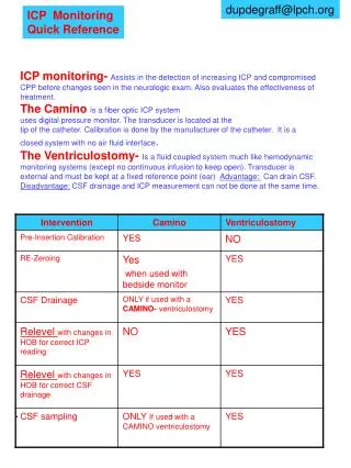
ICP Monitoring Techniques • Types of Monitoring Devices • Fluid-filled transduced ventriculostomy • Fiberoptic sensors • Microchips (internal strain-gauge devices) • Air pouch technologies • Locations of Monitoring Devices • Ventricular • Parenchymal • Subdural
Fluid-Filled Ventriculostomy with External Strain-Gauge Transducer • Fluid-filled transduced ventriculostomy • Consists of a catheter or hollow tube that is placed in the anterior horn of the lateral ventricle • Often tunneled under the scalp a short distance away from the burr hole insertion site before they exit the scalp; others are secured using a bolt • Tunneling under the scalp is thought to decrease infection • Catheter is attached to pressure resistant or “pressurized tubing,” filled with a preservative-free saline solution and connected to a nonflush strain-gauge transducer
Fluid-Filled Ventriculostomy with External Strain-Gauge Transducer • Transducer is leveled or zeroed at an external reference point that represents the level of the foramen of Monro • Methods used to reference the foramen of Monro: • Measuring halfway between the outer canthus of the eye and the tragus • Measuring one centimeter posterior to the eye or two centimeters above the pterion • Drawing a line between the two external auditory meatus and determining the midpoint • Using the external auditory meatus as the reference
Fluid-Filled Ventriculostomy with External Strain-Gauge Transducer • External strain-gauge transducers are connected to the fluid-filled ventriculostomies to measure the pressure transmitted from the CSF in the ventricles
Fluid-Filled Ventriculostomy with External Strain-Gauge Transducer • Typically measure the electrical resistance caused by a change in the strain present in a length of thin metal foil • Strain-gauge pressure sensing element sits atop a mechanical diaphragm • Ventricular catheter and pressure tubing conduct fluid and, therefore, intracranial pressure to the rear side of the diaphragm • Changes in intracranial pressure cause changes in the pressure exerted on the diaphragm and hence the strain on the sensor element • Simple electrical circuits are used to measure the resistance of the sensor element that is proportional to intracranial pressure
Transducer-Tipped Catheters • Primary pressure transducer is mounted on the distal tip of the implanted catheter • Fiberoptic and electrical (miniaturized strain-gauge) sensors are used in these devices
Transducer-Tipped Catheters • The primary sensor used is a mechanical diaphragm that moves with changes in pressure • Intensity Modulation (Camino) • Position of the diaphragm (and therefore pressure) alters the intensity of the light reflected from its rear surface • Interferometer • Position of the diaphragm is sensed by measuring the ratio of returned light intensities in 2 spectral bandwidths • Ratio is a function of spectral interference that varies with the position of the diaphragm
Transducer-Tipped Catheters • Strain-gauge device • Uses a miniaturized silicon strain-gauge located on the side of the catheter near the distal tip • Changes in the position of the diaphragm cause changes in the electrical resistance that is recorded and transformed and displayed as ICP readings • These systems may be used to measure pressure intraventricular, parenchymal, subdural, or epidural pressure
Air Pouch Technology • Senses intracranial pressure by filling a balloon that surrounds the end of the catheter with a set volume of air (0.05 to 0.1 cc) • Pressure exerted on the balloon pouch is the pressure of the surrounding tissue • Catheter re-zeros itself hourly by deflating and re-inflating the balloon to maintain a constant balloon volume
Compartmentalization of the Brain • Intracranial pressure can be monitored in several locations • Intraventricular, parenchymal, subdural, subarachnoid, or epidural • Accuracy varies • Pressures can vary • Within the intracranial compartment • The brain and CSF • Infratentorial and supratentorial • Between hemispheres • Because the contents are not homogenous, pressures vary even without pathology as a result of tissue and capillary density, though the differences may not be significant
Compartmentalization of the Brain • At the turn of the century, Harvey Cushing suggested that pressures were not evenly distributed throughout the brain, though to this day, clinicians continue to debate this • Researchers have looked at the phenomenon of compartmentalization for nearly 50 years with varying results
Ventriculostomy and CSF Drainage • Advantage of using a ventriculostomy to monitor ICP is that it can also be used to treat increased ICP by draining CSF • Cerebrospinal fluid plays a major role in the management of ICP • Believed that by reducing CSF volume, there is an increase in CBF and therefore an improvement in cerebral perfusion • Few studies have looked at the effectiveness of draining CSF on ICP and CBF
Ventriculostomy and CSF Drainage • Fortune J, Feustel P, Graca L, Hasselbarth J, Kuehler D. Effect of hyperventilation, mannitol, and ventriculostomy drainage on cerebral blood flow after head injury. J Trauma. 1995;39:1091–1099 • Examined the impact of • CSF drainage (open to drain for 3 minutes, drain set at level of foramen of Monro) • Mannitol (25 gms IV over 5 min) • Hyperventilation (decrease in PaCO2 about 5 mm Hg) • On • Increased ICP (ICP >15 mm Hg for 5 min) • CBF—using SjO2 as a surrogate for CBF
Ventriculostomy and CSF Drainage • Found that • Although all treatments lowered ICP, only mannitol improved CBF • CSF drainage had the most dramatic drop in ICP • Effect was often transient • They suggested that the magnitude and the duration that the ICP was controlled by the CSF drainage may be related to the intracranial compliance • If the intracranial compliance is decreased, small increases in volume result in large increases in pressure • Assessed by addition or removal of CSF and observation of the magnitude of the ICP change
Accuracy • A major concern for clinicians is the accuracy of ICP monitoring technology • ICP may be underestimated or overestimated • User must have an understanding of the limitations of the device being used • Compartmentalization within the cranium • Drift • Leveling of the transducer to obtain an accurate ICP measurement
Accuracy • Surface monitors, such as epidural and subdural catheters and subarachnoid bolts or screws, have been shown to be inaccurate • Thought that these devices are not representative of events occurring deep within the brain
Accuracy • Weinstabl et al found that Gaeltec epidural catheters recorded higher ICPs than the Camino subdural catheter • Believed that the subdural pressure was more representative of the true ICP • Inaccuracies in fluid-filled systems, such as subarachnoid bolts, arise from • Small leaks in the stopcocks • Improper positioning of the bolt (the tip must be located in the subarachnoid space) • Debris in the tip of the bolt impeding fluid pulsation Weinstabl C, Richling B, Plainer B, Czech T, Spiss C. Comparative analysis between epidural (Gaeltec) and subdural (Camino) intracranial pressure probes. J Clin Monitor. 1992;8:116–120.
Drift • Standards for ICP technology have been established by the Advancement of Medical Instrumentation • Include the following specification: • Pressure range measures between 0 to 100 mm Hg • Accuracy of ±2 mm Hg over a range of 0 to 20 mm Hg • Maximum error of 10% over a range of 20 to 100 mm Hg • Drift is defined as off set of zero and error in reading in response to changes in temperature
Drift Manufacturer' Zero Drift Specifications
Fluid-Filled Systems • Several key elements to obtaining an accurate measurement using a fluid-filled system • Include the in-situ catheter, low-compliant, unobstructed tubing and the appropriate transducer connection to the bedside monitor • Small, intraluminal size (<7F) of the in-situ catheter increases the frictional resistance of the CSF fluid and impacts pressure • The length, diameter, and the flexibility of the tubing used can alter the fidelity of the recording • Flexible, soft tubing (compliant), a large diameter tube, and excessive length in the tubing (>4 feet) dampen or blunt the pressure recording resulting in an underestimation the ICP • Debris in the catheter or tubing (brain matter, blood clots) and increased viscosity of the fluid (blood, infection, increased protein content) can also affect the reading
Fluid-Filled Systems • Air bubbles in the transducer, tubing, or stopcocks will dampen the pressure waveform, potentially leading to ICP measurement errors • The transducer must be leveled at the foramen of Monro and re-zeroed at regular intervals to maintain accuracy • Current transducers drift ± 2 mm Hg per 8 hours • If the transducer and extraventricular drain (EVD) are not referenced to the foramen of Monro correctly using a carpenter level's, bubble-line level, or laser level there can be a significant error • For every inch deviation above or below the reference, there is a 1.86 mm Hg error or 0.73 mm Hg for every centimeter • As the transducer falls below zero, the pressure increases and it decreases when it is raised above zero
Leveling • Study of leveling practices at a 450-bed academic medical center • Visual checks of the level of the transducer/drainage chamber resulted in a mean error of 4.4 centimeters (3.2 mm Hg) • Error lessened when a carpenter's level was used (mean 1.3 centimeters, 0.95 mm Hg) • Better with the use of a laser level device (mean 0.9 centimeters of water, 0.7 mm Hg • Brisnaire D, Robinson L. Accuracy of leveling intraventricular collection drainage systems. J Neurosci Nurs. 1997;29:261–268.
Leveling • Also found that 5 out of 33 participants did not know the proper reference for the foramen of Monro • An imaginary line drawn between the top of the ear and outer canthus of the eye • 9 of 33 participants did not know how to properly adjust the drainage chamber • Years of experience in the intensive care unit (>5 years) and participation in in-service education (50% attendance) appear to influence accuracy
Complications • Infection • Hemorrhage • Breakage • Malfunction of the device • Difficulty with placement • Difficult to assess the true rate of some of these issues • Variability of definitions used in the literature when reporting infections and hemorrhage
Infections • May be defined as: • A positive cerebrospinal fluid (CSF) culture obtained from ventricular or lumbar catheter • Positive culture with CSF pleocytosis, low glucose level, or high protein level • CSF pleocytosis or low glucose level alone without a positive culture • Presence of clinical symptoms as fever or mental status changes • 2 positive cultures with same organisms
Infections • Contamination • An isolated CSF culture with normal CSF cell counts and absent clinical symptoms • Or a negative-Gram stain with a positive culture • Difficult to ascertain the incidence of CNS infections resulting from ICP monitoring devices because of the variability in what is defined as an infection • Overall infection rates regardless of definition range from 0% to 27%; many of these had poor or no definition of infection
Infections • Infections rates for tunneled ventricular catheters appear to be between 0% and 4% though data is not well defined • Reported infection rates for intraparenchymal devices ranged from 0.3% to 3.7% • One study reported a 7% incidence of positive tip culture from intraparenchymal devices but no evidence of clinical infection
Infections • Time the device was indwelling (greater than 5 days) and location in the hospital the catheter was inserted (outside of operating room) correlate with higher infection rates • Using strict aseptic technique during insertion (gown, glove, mask) and when manipulating the device is essential to prevent contamination • Most commonly reported pathogens are Staphylococcus aureus and epidermis, E coli, Klebsiella, and Streptococcus • The benefit of prophylactic antibiotics is unclear
Infections in VP Shunts • The organisms involved and the route of infection vary with the time of onset of the infection • Most infections caused by coagulase-negative staphylococci occur shortly after placement of the shunt (eg, 50 percent within two weeks and 79 percent within two months in two series) • The organisms are most often introduced at the time of the procedure from the patient's own skin flora, operative personnel, or the environment • S. aureus is also a common cause of early onset shunt infection
Infections in VP Shunts • Infections occurring more than nine months after shunt surgery account for 12 to 13 percent of shunt infections and are often caused by organisms other than staphylococci • In a review of 40 such infections • 18 infections were due to peritonitis, most often arising from appendicitis or bowel perforation • 8 were due to hematogenous seeding • 4 due to direct inoculation after abdominal surgery or traumatic exposure of the shunt • In 10 no identifiable cause was identified
Infections in VP Shunts • Bacteria involved were: • Intestinal flora (in association with peritonitis) • Bacteremia organisms (eg, Streptococcus pneumoniae, Listeria monocytogenes) • Skin flora (introduced at the time of shunt placement or from surgery or traumatic exposure at a later date)
Infections in VP Shunts • Does the shunt need to be tapped? • Direct aspiration of the shunt is preferred and yields a positive culture in more than 90% of cases • By contrast, CSF obtained by lumbar puncture or ventricular tap in one study was culture-positive in only 58 and 79% of cases for VA and VP shunts, respectively
Hemorrhage • Implantation of any device in the brain carries a potential risk of bleeding • Proper training on insertion procedures can minimize these risks • Hemorrhages are often not reported or may be clinically insignificant
Hemorrhage • In recent studies, attempts have been made to classify the significance of the hemorrhages • Blaha et al defined hemorrhages in 3 grades: • Grade 1: small punctuate hemorrhage or local SAH • Grade 2: ICH, diffuse SAH or extra axial hematoma without new neurologic deficit requiring craniotomy • Grade 3: ICH, diffuse SAH or extra-axial hematoma with new neurologic deficit requiring craniotomy
Hemorrhage • They reported an overall hemorrhage rate of 9.2% with intraparenchymal devices: • 7.5% were grade 1 • 2.2% were grade 2 • Anderson et al reported a 6.4% incidence of hemorrhage for intraparenchymal devices: • 4.8% were grade 1 • 1.6% were grade 2 • Neither study reported grade 3 bleeds • Anderson et al reported a 17.6% incidence of hemorrhage with intraventricular catheters with one bleed requiring surgical intervention
Hemorrhage • Role of coagulopathy as a contributing factor in hemorrhage is often a concern • particularly with acute trauma and liver failure • Recommended the coagulapathy be corrected before placing an ICP monitor • Davis et al • Risk of hemorrhage in patients with borderline International Normalized Ratio (INR) of 1.3 to 1.6 showed no increased risk of bleeding and therefore no benefit in continued infusion of fresh frozen plasma to correct the INR
Hemorrhage • Correcting coagulation factors is a difficult issue in patients with fulmination hepatic failure • These patients may have quite high ICPs • Some physicians are reluctant to place intraparenchymal catheters because of the risk of bleeding and, as a result, they may use the less reliable surface monitors • Some publications demonstrate the safe use of not only intraparenchymal ICP catheters but microdialysis in this population
Other Complications • Other concerns when placing an ICP monitors are: • Device malfunction • Catheter malfunction or breakage is primarily an issue of advanced technology • Improper or difficulty with placement • Most frequently seen with ventricular catheters because the ventricles are shifted laterally off midline or slit ventricles • Dislodging of the device
Other Complications • Concerns over infection rates, hemorrhage, malposition, breakage, and dislodgement can be addressed by proper training, practice, and taking care when placing and using the technology • Following the manufacturer's recommendations for insertion will limit both trauma to the patient from the catheter insertion and damage to catheters and will ensure proper placement of the device and, therefore, accurate ICP recordings • Securing the catheters after insertion and using care when moving patients will prevent dislodgement and breakage of these devices