Download
1 / 103

E N D
Pathognomonic clinical signs of traumatic tissue maxillofacial area in children. Modern diagnostic methods. The principles of therapeutic tactics in injuries of the soft tissues of the face, teeth, bones. Diagnosis, differential diagnosis and treatment of TMJ ankilosis. Modern principles of treatment and rehabilitation of children with congenital maxillo facial area.
Maxillofacial trauma Management of traumatized patient
Causes: △ Road traffic accident (RTA) 35-60% Rowe and Killey 1968; Vincent-Towned and Shepherd 1994 △ Fight and assault (interpersonal violence) Most in economically prosperous countries Beek and Merkx 1999 △ Sport and athletic injuries △ Industrial accidents △ Domestic injuries and falls
Incidence Literatures reported different incidence in different parts of the WORLD and at different TIMES √ 11% in RTA (Oikarinen and Lindqvist 1975) Mandible (61%) Maxilla (46%) Zygoma (27%) Nasal (19.5%)
Factors affecting the high/low incidence of maxillofacial trauma Geography Fight, gunshot and RTA in developed and developing countries respectively (Papavassiliou 1990, Champion et al 1997) Social factors Violence in urban states (Telfer et al 1991; Hussain et al 1994; Simpson & McLean 1995) Alcohol and drugs Yong men involved in RTA wile they are under alcohol or drug effects (Shepherd 1994) Road traffic legislation Seat belts have resulted in dramatic decrease in injury (Thomas 1990, as reflected in reduction in facial injury (Sabey et al 1977) Season Seasonal variation in temperature zones (summer and snow and ice in midwinter) of RTA, violence and sporting injuries (Hill et al 1998)
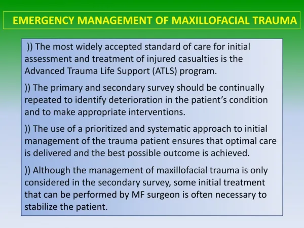
Assessment of traumatized patient This should not concentrate on the most obvious injury but involve a rapid survey of the vital function to allow management priorities 5% of all deaths world wide are caused by trauma This might be much higher in this country
Peaks of mortality First peak Occurs within seconds of injury as a result of irreversible brain or major vascular damage Second peak Occurs between a few minutes after injury and about one hour later (golden hour) Third peak Occurs some days or weeks after injury as a result of multi-organ failure
Organization of trauma services Pre-hospital care (field triage) Care delivered by fully trained paramedic in maintaining airway, controlling cervical spine, securing intravenous and initiating fluid resuscitation Hospital care (inter-hospital triage) Senior medical staff organized team to ensure that medical resources are deployed to maximum overall benefit Mass casualty triage triage decisions are crucial in determining individual patients survival
Primary survey ⒶAirway maintenance with cervical spine control Ⓑ Breathing and ventilation ⒸCirculation with hemorrhage control ⒹDisability assessment of neurological status Ⓔ Exposure and complete examination of the patient
Airway Satisfactory airway signifies the implication of breathing and ventilation and cerebral function Management of maxillofacial trauma is an integral part in securing an unobstructed airway Immobilization in a natural position by a semi-rigid collar until damaged spine is excluded
Sequel of facial injury Obstruction of airway asphyxia Cerebral hypoxia Brain damage/ death Is the patient fully conscious? And able to maintain adequate airway? Semiconscious or unconscious patient rapidly suffocate because of inability to cough and adopt a posture that held tongue forward
Immediate treatment of airway obstruction in facial injured patient △Clearing of blood clot and mucous of the mouth and nares and head position that lead to escape of secretions (sit-up or side position) △ Removal of foreign bodies as a broken denture or avulsed teeth which can be inhaled and ensuring the patency of the mouth and oropharynex △ Controlling the tongue position in case of symphesial bilateral fracture of mandible and when voluntary control of intrinsic musculature is lost △ Maintaining airway using artificial airway in unconscious patient with maxillary fracture or by nasophryngeal tube with periodic aspiration △ Lubrication of patient’s lips and continuous supervision
Additional methods in preservation of the airway in patient with severe facial injuries Endotracheal intubation Needed with multiple injuries, extensive soft tissue destruction and for serious injury that require artificial ventilation Tracheostomy Surgical establishment of an opening into the trachea Indications: 1. when prolonged artificial ventilation is necessary 2. to facilitate anesthesia for surgical repair in certain cases 3. to ensure a safe postoperative recovery after extensive surgery 4. following obstruction of the airway from laryngeal edema 5. in case of serious hemorrhage in the airway Circothyroidectomy An old technique associated with the risk of subglottic stenosis development particularly in children. The use of percutaneous dilational treachestomy (PDT) in MFS is advocated by Ward Booth et al (1989) but it can be replaced with PDT. Control of hemorrhage and Soft tissue laceration Repair, ligation, reduction of fracture and Postnasal pack
Cervical spine injury Can be deadly if it involved the odontoid process of the axis bone of the axis vertebra If the injury above the clavicle bone, clavicle collar should minimize the risk of any deterioration
Breathing and ventilation Chest injuries: Pneumothorax, haemopneumothorax, flail segments, reputure daiphram, cardiac tamponade signs Clinical Deviated trachea Absence of breath sounds Dullness to percussion Paradoxical movements Hyper-response with a large pneumothorax Muffled heart sounds Radiographical Loss of lung marking Deviation of trachea Raised hemi-diaphragm Fluid levels Fracture of ribs
Emergency treatment in case of chest injury Occluding of open chest wounds Endotreacheal intubation for unstable flail chest Intermittent positive pressure ventilation Needle decompression of the pericardium Decompression of gastric dilation and aspiration of stomach content
Circulation Circulatory collapse leads to low blood pressure, increasing pulse rate and diminished capillary filling at the periphery Patient resuscitation Restoration of cardio-respiratory function Shock management Replacement of lost fluid
Fluid for resuscitation: ☞Adequate venous access at two points ☞ Hypotension assumed to be due to hypovolaemia ☞ Resuscitation fluid can be crystalloid, colloid or blood; ringer lactate ☞ Surgical shock requires blood transfusion, preferably with cross matching or group O+ ☞ Urine output must be monitored as an indicator of cardiac out put
Reduction and fixation will often arrest bleeding of long duration Pulse and blood pressure should be monitored and appropriate replacement therapy is to be started
Neurological deficient Rapid assessment of neurological disability is made by noting the patient response on four points scale: A Response appropriately, is Aware V Response to verbal stimuli P Response to painful stimuli U Does not responds, Unconscious
Glasgow coma scale (GCS)(Teasdale and Jennett, 1974) Score 8 or less indicates poor prognosis, moderate head injury between 9-12 and mild refereed to 13-15
Exposure All trauma patient must be fully exposed in a warm environment to disclose any other hidden injuries When the airway is adequately secured the second survey of the whole body is to be carried out for: Accurate diagnosis Maintenance of a stable state Determination of priorities in treatment Appropriate specialist referral
Secondary surveyAlthough maxillofacial injuries is part of the secondary survey, OMFS might be involved at early stage if the airway is compromised by direct facial trauma Head injury Abdominal injury Injury to extremities
Head injuryMany of facial injury patients sustain head injury in particular the mid face injuries Open Closed it is ranged from Mild concussion to brain death
Signs and symptoms of head injury Loss of conscious OR History of loss of conscious History of vomiting Change in pulse rate, blood pressure and pupil reaction to light in association with increased intracranial pressure Assessment of head injury (behavioral responses “motor and verbal responses” and eye opening) Skull fracture Skull base fracture (battle’s sign) Temporal/ frontal bone fracture Naso-orbital ethmoidal fracture
slow reaction and fixation of dilated pupil denotes a rise in intra-cranial pressure Rise in intercranial pressure as a result of acute subdural or extradural hemorrhage deteriorate the patient’s neurological status Apparently stable patient with suspicion of head injury must be monitored at intervals up to one hour for 24 hour after the trauma
Hemorrhage Acute bleeding may lead to hemorrhagic shock and circulatory collapse Abdominal and pelvis injury; liver and internal organs injury (peritonism) Fracture of the extremities (femur)
Abdomen and pelvis In addition to direct injuries, loss of circulating blood into peritoneal cavity or retroperitonial space is life threatening, indicated by physical signs and palpation, percussion and auscultation Management: Diagnostic peritoneal lavage (DPL) to detect blood, bowel content, urine Emergency laprotomy
Extremity trauma Fracture of extremities in particular the femur can be a significant cause of occult blood loss. Straightening and reduction of gross deformity is part of circulation control Cardinal features of extremities injury Impaired distal perfusion (risk of ischemia) Compartment syndrome (limb loss) Traumatic amputation
Patient hospitalization and determination of prioritiesFacial bone fracture is hardly ever an urgent procedure,simple and minor injury of ambulant patient may occasionally mask a serious injury that eventually ended the patient’s life △ emergency cases require instant admission △ conditions that may progress to emergency △ cases with no urgency
Preliminary treatment in complex facial injury Soft tissue laceration (8 hours of injury with no delay beyond 24 hours) Support of the bone fragments Injury to the eye As a result of trauma, 1.6 million are blind, 2.3 million are suffering serious bilateral visual impairment and 19 million with unilateral loss of sight (Macewen 1999) Ocular damage Reduction in visual acuity Eyelid injury
Prevention of infectionFractures of jaw involving teeth bearing areas are compound in nature and midface fracture may go high, leading to CSF leaks (rhinorrhoea, otorrhoea) and risk of meningitis,and in case of perforation of cartilaginous auditory canal Diagnosis: Laboratory investigation, CT and MRI scan Management: Dressing of external wounds Closure of open wounds Reposition and immobilization of the fractures Repair of the dura matter Antibacterial prophylaxis (as part of the general management (Eljamal, 1993)
Control of painDisplaced fracture may cause severe pain but strong analgesic ( Morphine and its derivatives) must be avoided as they depress cough reflex, constrict pupils as they may mask the signs of increasing intracranial pressure Management: ☞ Non-steroidal anti-inflammatory drugs can be prescribed (Diclofenac acid) ☞ Reduction of fracture ☞ sedation
In patient care Necessary medications Diet (fluid, semi-fluid and solid food) intake and output (fluid balance chart) Hygiene and physiotherapy Proper timing for surgical intervention
Pathophysiology Maxillofacial fractures result from either blunt or penetrating trauma. Penetrating injuries are more common in city hospitals. Midfacial and zygomatic injuries. Blunt injuries are more frequently seen in community hospitals. Nose and mandibular injuries.
Pathophysiology High Impact: Supraorbital rim – 200 G Symphysis of the Mandible –100 G Frontal – 100 G Angle of the mandible – 70 G Low Impact: Zygoma – 50 G Nasal bone – 30 G
Etiology @60% of patients with severe facial trauma have multisystem trauma and the potential for airway compromise. 20-50% concurrent brain injury. 1-4% cervical spine injuries. Blindness occurs in 0.5-3%
Etiology 25% of women with facial trauma are victims of domestic violence. Increases to 30% if an orbital wall fx is present. 25% of patients with severe facial trauma will develop Post Traumatic Stress Disorder
Emergency ManagementAirway Control Control airway: Chin lift. Jaw thrust. Oropharyngeal suctioning. Manually move the tongue forward. Maintain cervical immobilization
Emergency ManagementIntubation Considerations Avoid nasotracheal intubation: Nasocranial intubation Nasal hemorrhage Avoid Rapid Sequence Intubation: Failure to intubate or ventilate. Consider an awake intubation. Sedate with benzodiazepines.
Emergency ManagementIntubation Considerations Consider fiberoptic intubation if available. Alternatives include percutaneous transtracheal ventilation and retrograde intubation. Be prepared for cricothyroidotomy.
Emergency ManagementHemorrhage Control Maxillofacial bleeding: Direct pressure. Avoid blind clamping in wounds. Nasal bleeding: Direct pressure. Anterior and posterior packing. Pharyngeal bleeding: Packing of the pharynx around ET tube.
History Obtain a history from the patient, witnesses and or EMS. AMPLE history Specific Questions: Was there LOC? If so, how long? How is your vision? Hearing problems?
History Specific Questions: Is there pain with eye movement? Are there areas of numbness or tingling on your face? Is the patient able to bite down without any pain? Is there pain with moving the jaw?
Physical Examination Inspection of the face for asymmetry. Inspect open wounds for foreign bodies. Palpate the entire face. Supraorbital and Infraorbital rim Zygomatic-frontal suture Zygomatic arches
Physical Examination Inspect the nose for asymmetry, telecanthus, widening of the nasal bridge. Inspect nasal septum for septal hematoma, CSF or blood. Palpate nose for crepitus, deformity and subcutaneous air. Palpate the zygoma along its arch and its articulations with the maxilla, frontal and temporal bone.
Physical Examination Check facial stability. Inspect the teeth for malocclusions, bleeding and step-off. Intraoral examination: Manipulation of each tooth. Check for lacerations. Stress the mandible. Tongue blade test. Palpate the mandible for tenderness, swelling and step-off.
Physical Examination Check visual acuity. Check pupils for roundness and reactivity. Examine the eyelids for lacerations. Test extra ocular muscles. Palpate around the entire orbits..